Abstract
This review of kinaesthesia, the senses of limb position and limb movement, has been prompted by recent new observations on the role of motor commands in position sense. They make it necessary to reassess the present-day views of the underlying neural mechanisms. Peripheral receptors which contribute to kinaesthesia are muscle spindles and skin stretch receptors. Joint receptors do not appear to play a major role at most joints. The evidence supports the existence of two separate senses, the sense of limb position and the sense of limb movement. Receptors such as muscle spindle primary endings are able to contribute to both senses. While limb position and movement can be signalled by both skin and muscle receptors, new evidence has shown that if limb muscles are contracting, an additional cue is provided by centrally generated motor command signals. Observations using neuroimaging techniques indicate the involvement of both the cerebellum and parietal cortex in a multi-sensory comparison, involving operation of a forward model between the feedback during a movement and its expected profile, based on past experience. Involvement of motor command signals in kinaesthesia has implications for interpretations of certain clinical conditions.
The term ‘kinaesthesia’ was coined by Bastian (1888) and refers to the ability to sense the position and movement of our limbs and trunk. It is a mysterious sense since, by comparison with our other senses such as vision and hearing, we are largely unaware of it in our daily activities. In the absence of vision we know where our limbs are but there is no clearly defined sensation that we can identify. In this review we are reporting a number of recent developments in the field that promise to redefine it. They concern the role of motor commands in limb position sense. The debate about a ‘sensation of innervations’ has raged since the 19th century and the new findings discussed here finally lay any doubts to rest. The subject of kinaesthesia is steeped in history and readers interested in the historical background should consult Goodwin et al. (1972), McCloskey (1978) and Donaldson (2000). For more recent reviews see Gandevia (1996) and Proske (2005, 2006).
The receptors
The intuitively obvious place to look for receptors subserving the sense of joint position and movement is in the joints themselves and that is what was believed for much of the 20th century (e.g. Mountcastle & Powell, 1959; Skoglund, 1973). The ground-breaking observations of Goodwin et al. (1972) demonstrated the importance for kinaesthesia of receptors located in the muscles rather than in joints, in particular, the role played by muscle spindles. Today it is accepted that muscle spindles make a major contribution. In addition, receptors from skin and signals of central origin associated with the sense of effort or will and derived from the motor command, have become recognised as playing important roles.
The principal muscle receptor in kinaesthesia is the muscle spindle. It includes both the primary and secondary endings of spindles. Primary endings respond to the size of a muscle length change and its speed (Matthews, 1972). They are therefore believed to contribute both to the sense of limb position and movement (see below). Secondary endings do not have a pronounced velocity sensitivity and signal only the length change itself, so contribute only to the sense of position. For more detailed discussions see Matthews (1972).
At the time of the experiments of Goodwin et al. (1972) it was already known that muscle spindles in animals, specifically the primary endings, were exquisitely sensitive to muscle vibration (Brown et al. 1967). It was shown subsequently that this applied also to human spindles (e.g. Burke et al. 1976; Roll et al. 1989). Goodwin and colleagues showed that vibration of elbow muscles produced illusions of both forearm movement and displacement during the period of vibration. Importantly, when the vibrator was moved from the muscle belly to the elbow joint, the illusion disappeared.
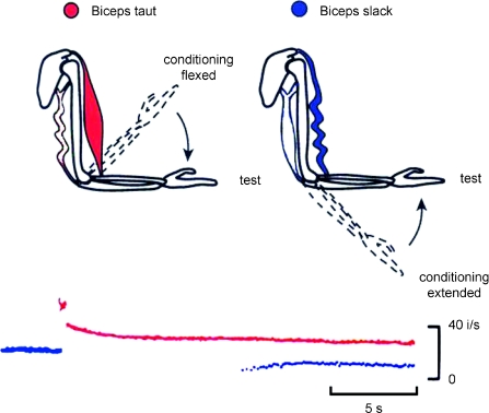
Evidence from a rather different source, supporting the view of a significant contribution from muscle spindles to kinaesthesia comes from the dependence of passive tension in muscle, both extrafusal and intrafusal, on the previous history of contraction and length changes, muscle thixotropy (Proske et al. 1993). In resting muscle, long-lasting stable cross-bridges develop between actin and myosin (Hill, 1968; Proske & Morgan, 1999). This gives passive muscle fibres a degree of stiffness and they fall slack if subsequently shortened. For muscle spindles it means that background discharge rates change, depending on what has happened to the muscle beforehand. If slack is present, discharge rates are low, if slack has been removed by a contraction, they can be increased severalfold (Gregory et al. 1988). Changes in muscle history, called conditioning, can lead to errors in limb position sense (Gregory et al. 1988; Winter et al. 2005; see Fig. 1).
Figure 1. The technique of muscle conditioning.
The two diagrams at the top show a human forearm with one flexor and one extensor muscle drawn in, the flexor being colour-coded. On the left, the arm is held flexed (dashed lines) and the flexors are contracted (conditioning flexed). Once the arm has relaxed it is moved to an intermediate angle (test). This leaves biceps and its spindles in a ‘taut’ state (red). When the arm is held extended (dashed lines) and elbow extensors are contracted (conditioning extended), moving the arm to the intermediate angle (test) leads to development of ‘slack’ in biceps and its spindles (blue). The lower diagram shows an instantaneous frequency display of the responses of a muscle spindle in the cat soleus muscle following conditioning of the muscle leaving the spindle taut (red) or slack (blue). Muscle conditioning leads to substantial changes in spindle discharge rates and that, in turn, leads to errors in forearm position sense. Redrawn, in part, from Wood et al. (1996).
Goodwin et al. (1972) found that the kinaesthetic illusion from vibration was predominantly one of movement, although there was a perceived change in limb position during and at the end of vibration. Muscle conditioning, on the other hand, produces changes in perceived position only, as a result of altered background firing rates in muscle spindles. It has recently been shown in a two-arm position-matching task that conditioning one arm by contracting its elbow flexors while the arm is held flexed and conditioning the other by contracting its elbow extensors with the arm held extended, led to a 20 deg error in position matching (Allen et al. 2007; White & Proske, 2009). On interrogation the blindfolded subjects insisted that their arms were accurately aligned. Such a result emphasises the powerful influence exerted by muscle history effects on position sense. Only muscle spindles would be expected to show such behaviour, dramatically altering their signalling properties as a result of a muscle contraction.
Studies using other techniques such as skin and joint anaesthesia and the effective disengagement of muscles from joints also point to the importance of the role of muscle receptors in the detection of passive movements. For a review, see Gandevia (1996).
Concerning the possible contribution to kinaesthesia from other receptor types, the summary view is that while a good case has been made for some cutaneous receptors, the evidence is less convincing for joint receptors. The cutaneous receptor most likely to subserve a kinaesthetic role is the skin stretch receptor, the slowly adapting Type II receptor served by Ruffini endings (Chambers et al. 1972; Edin, 1992). For kinaesthesia at the forearm, stretch of skin over the elbow during elbow flexion can provide information about both position and movement. Movement illusions generated by stretch of skin of the hand and over more proximal joints, when combined with muscle vibration were greater than when either stimulus was applied on its own (Collins et al. 2005). The authors made the point that this was not just a matter of skin input facilitating the muscle input and that cutaneous input generated by skin stretch contributed to kinaesthesia in its own right. More recent observations have shown that skin input can also have an occluding action. Signals from local, rapidly adapting receptors evoked by low-amplitude, high frequency vibration can impede movement detection (Weerakkody et al. 2007).
An example of a situation where skin receptors play an indispensable role in kinaesthesia is in skin adjacent to the finger joints. The muscles that move the fingers lie in the forearm and hand and their tendons cross more than one joint. In this situation the muscle afferent information is potentially ambiguous. The proximity of skin receptors adjacent to each joint allows them to provide joint-specific information (Collins et al. 2005). Furthermore, it has recently been pointed out that whenever a muscle spans more than one joint this can compromise its spindles’ ability to detect movements (Sturnieks et al. 2007).
While joint receptors were first thought to be all-important in kinaesthesia, the present-day view is that their contribution at most joints is likely to be minor. Typically they respond to joint movement, but often with response peaks at both limits of the range of joint motion (Burgess & Clark, 1969). They are now thought of as limit detectors. However, there are examples in the literature of responses across the full range of joint movement (Burke et al. 1988) and here joint receptors may play a role under circumstances in which input from muscle and skin is not available (Ferrell et al. 1987).
Two senses
An important question is whether the senses of position and movement should be considered part of the same single sense or as two separate entities. It will be agued here that the two are separate, although they obviously have some elements in common.
The belief that the two comprise part of a single sense is due partly to historical reasons (Bastian, 1888) and because they share common receptors of origin. As already stated, primary endings of spindles provide information about both position and movement. Consistent with this is the observation that if movements are made progressively more slowly, what started out as a movement sensation eventually blends into a sense of changed position (Clark et al. 1985; Taylor & McCloskey, 1990). Similarly, muscle conditioning produces an alteration of spindle background firing rates and this is accompanied by changes in limb position sense (Gregory et al. 1988).
In a study of the effects of muscle vibration, McCloskey (1973) observed that when the frequency of vibration was reduced from 100 Hz to 20 Hz, the sensation of movement and changed position gradually blended into one of position only. Presumably at the lower frequency the input included a component from spindle secondary endings which would be insensitive to the higher frequencies. McCloskey argued that the two senses were generated by separate lines of input and that position sense was not derived from an integration of the velocity signal (see also Sittig et al. 1985).
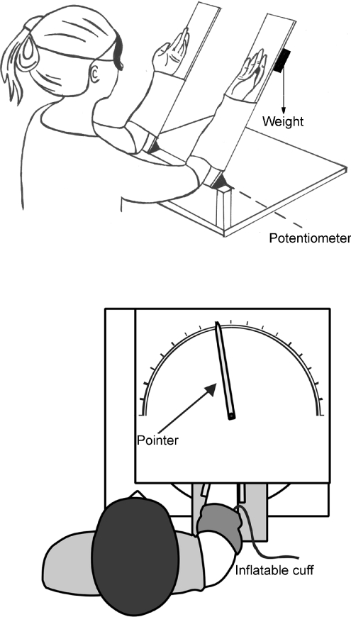
Another piece of evidence comes from the effects of fatigue on kinaesthesia (Allen & Proske, 2006). Position matching at the elbow joint was measured by placing one forearm at a given angle and asking blindfolded subjects to match its position with the other arm (Fig. 4). Movement sense was measured by asking subjects to track with their other arm movements at different speeds imposed on one arm. After exercise that reduced maximal elbow flexor force by 30%, significant errors had developed in position sense, but not in movement sense.
Figure 4. Measuring position sense.
Upper panel, apparatus used for position matching at the elbow joint. The experimenter places one arm at a set angle and the blindfolded subject matches its position with their other arm. The arms are supported by a pair of paddles which are hinged at a point aligned with the elbow joint and potentiometers at the hinge give the elbow angle. A weight can be attached under the paddle to load the arm. (Redrawn from Allen & Proske, 2006, with kind permission of Springer Science + Business Media.) Lower panel, apparatus used to measure position of the hand at the wrist. The fingers are held in full extension by a pair of plates which can be rotated through a range of angles. At each angle the subject indicates the perceived position of their hand by moving the pointer with their other hand. A nerve block can be effected by inflation of a cuff on the upper arm to produce ischaemic paralysis and anaesthesia of the hand. A similar cuff is used to restrict the distribution of a paralysing drug injected into the lower arm. From Smith et al. 2009.
Observations on neurones of the primary motor cortex of awake monkeys indicate the presence of some neurones that code for load only during postural tasks, others that code for load only during movements and a third group that respond during both conditions (Kurtzer et al. 2005). It was concluded that distinct mechanisms underlie cortical processing of position-related activity. Graziano et al. (2002) described different postures adopted by monkeys during repetitive microstimulation at specific sites in primary motor cortex. The data suggested that positional information was a parameter of importance for the brain and its derivation was probably distinct from that for movement information. Finally, in a different experiment, blindfolded human subjects were required to move their arm between two targets for 70 repetitions. It was found that during the repeated movements hand position drifted without comparable drift in the distance or direction moved (Brown et al. 2003). This was interpreted as implying the existence of separate position and movement controllers. To conclude, while some receptors are able to contribute input to both the sense of position and the sense of movement, the balance of evidence suggests that the central processing of the information is done separately.
The sense of limb position
The neural basis of limb position sense is the ability of receptors like muscle spindles to maintain static levels of discharge which increase in proportion to the increase in muscle length. This has been shown for cat spindles (e.g. Bessou & Laporte, 1962; Proske et al. 1993) and human spindles (e.g. Roll & Vedel, 1982). Similar behaviour can be demonstrated for slowly adapting cutaneous receptors contributing to position sense (e.g. Edin, 1992).
As already mentioned, measurements of position sense in a relaxed limb depend on how the muscles of the limb were conditioned beforehand An effective way to condition the muscle is by contracting it isometrically with a 20% of maximum voluntary contraction (Gregory et al. 1998). The contraction will remove any slack in intrafusal fibres and raise spindle resting activity. Another method of conditioning is to use repeated, large-amplitude ‘stirring’ movements (Lakie & Robson, 1988). This leaves spindles in a less well-defined state. The stirring leads to development of slack in intrafusal and extrafusal muscle fibres. So if the influence on position sense of an isometric contraction is sought, any outcome may include a component due to the spindles signalling the take-up of slack (e.g. Winter et al. 2005).
If it is accepted that spindles play a major role in position sense, then an important problem relates to their position-signalling capacity during muscle contractions. Whenever we generate a voluntary contraction, both skeletomotor and fusimotor neurones are coactivated (Vallbo, 1971, 1974). When a limb is supporting a load, for example its own weight against gravity, the spindle signal from the agonist muscle will have changed as a result of coactivation. An important question for kinaesthesia is, how does the brain interpret the additional spindle activity generated through the fusimotor system?McCloskey et al. (1983) suggested that any fusimotor-evoked spindle signals are subtracted centrally. In jargon terms, the ‘reafferent’ signal would be subtracted, leaving only the ‘exafferent’ component to signal position.
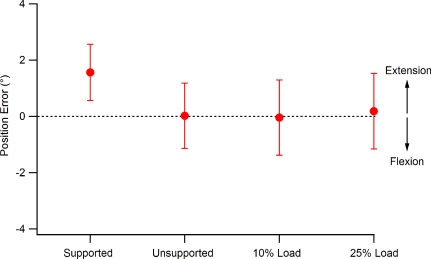
Whatever process the brain uses to overcome this difficulty, it is effective (Fig. 2). When blindfolded subjects matched the positions of their forearms, they were able to do so quite accurately, even when one arm was required to support a load of 25% of maximum (Allen et al. 2007). So despite the presence of significant motor activity in the muscles of one arm, position-matching ability remained unaffected. In the search for an explanation, rather than postulating a simple subtraction process, it has been suggested that a central copy of the motor command is used to access memories of similar movements to compute the expected feedback. This is compared with the actual feedback (Matthews, 1988). Similar mechanisms using a forward model have been treated more formally (Wolpert et al. 1995; Kawato, 1999; Cullen, 2004). In the situation where one arm is supporting a load, a contribution to the matching process may be provided by the unloaded matching arm. The signal from the loaded arm, where the exafference is submerged in a volume of reafference, may contribute less to the subject's decision of having achieved a satisfactory match. It is known that in a matching task signals from both arms are compared and their difference is used in placement of the indicator arm (Allen et al. 2007; White & Proske, 2009). Such a comparison would, of course, not be available when the task involved measurement of position sense in only one limb (Gandevia et al. 2006 and Fig. 4).
Figure 2. Position-matching errors in the vertical plane.
Values are means (±s.e.m.) for 12 subjects. Matching errors in the direction of extension are shown as positive. Dotted line, zero error. For this experiment both arms had been flexion conditioned before each matching trial. The four conditions for the reference arm were: supported by the experimenter, supported by the subject, supported by the subject with a 10% maximum voluntary contraction load added, and supported by the subject with a 25% load added. Despite one arm bearing an increasingly heavy load, matching accuracy remains unaffected. From Allen et al. (2007).
The sense of effort
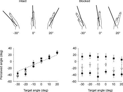
It has been known for some time that the sense of effort can contribute to force and heaviness sensations (e.g. Carson et al. 2002; Weerakkody et al. 2003). More recently it has been proposed that the sense of effort may also contribute to kinaesthesia. This is against a background of observations which did not support such a claim (Goodwin et al. 1972; McCloskey & Torda, 1975). It was believed that signals of motor command could calibrate sensory input coming from the periphery but did not generate sensations in their own right. For a review see Gandevia (1987). In these new experiments (Gandevia et al. 2006) conduction in both afferent and motor fibres to the hand was blocked with a pressure cuff applied to the upper arm. When subjects tried to move their paralysed, anaesthetised hand they perceived a distinct displacement of the hand by up to 20 deg (Fig. 3). This put beyond doubt the availability of an effort-related signal under conditions where no peripheral signals were available. It led to the question, were such signals still detectable when the full complement of afferent feedback was present? In a subsequent experiment (Smith et al. 2009), subjects’ hand muscles were paralysed with a neuromuscular blocker, but not anaesthetised. The paralysis was considered deep enough to block both extrafusal and intrafusal motor junctions. Here the hypothesis was that effort signals might only contribute to kinaesthesia in the absence of any reafference. During paralysis attempted movements with a 30% of maximum effort led to perceived hand displacements of 8–9 deg. Therefore, with afferents intact, the displacement illusion generated by isometric efforts was smaller than with a total nerve block but it was still present. The observation supported the view that central command signals contributed to kinaesthesia, even when afferent feedback was available.
Figure 3. Change in perceived position of a phantom hand during flexion and extension.
Left-hand panel, data before a total nerve block; right-hand panel, after the nerve block. Drawings at the top, when the relaxed hand was placed at each angular position, its perceived position is indicated by the black bar. Lower panel, perceived position of the relaxed hand (mean ±s.e.m.) at each of 6 angular positions (open triangles) and the perceived position during isometric efforts at 30% of maximum in the direction of flexion (filled circles) and the direction of extension (filled squares). Dashed line indicates accurate match. Before the nerve block, subjects are able to accurately indicate the position of the unseen hand. Isometric efforts produce only small errors. After the block, subjects are no longer able to indicate the position of the hand and efforts to move the hand produce large illusions of hand displacement. From Gandevia et al. (2006).
An important additional observation made by Smith et al. (2009) was that in the normal hand isometric flexion or extension contractions led to perceived displacements of the hand by 6–7 deg. It implied that during normal, active movements effort signals were always available to contribute to position sense. Such a conclusion is hard to reconcile with the observations on arm-matching accuracy when one arm is loaded (Figs 2 and 4). However, as mentioned earlier, in an arm-matching task the unloaded arm may be providing a position cue. Such a cue is not available when position sense is measured in only one limb. In the one-hand experiments it might be interesting in the future to try to manipulate the sense of effort by fatiguing hand muscles. This might be expected to increase the effort required to generate a given level of force and therefore increase the accompanying position errors. It is known that the effect of motor commands on position sense increases with effort (Gandevia et al. 2006; Smith et al. 2009). In any case, the evidence for a contribution to kinaesthesia from a centrally generated effort signal is compelling. Exactly how the mix of peripheral and central signals is combined during normal movements remains to be determined.
Central projection
Clinical medicine has provided some information about the site of origin and central projection of kinaesthetic information. Thus most kinaesthetic afferents must reside in muscle or skin since joint replacement surgery does not lead to any deficit in kinaesthesia (Grigg et al. 1973). It is commonly believed that all kinaesthetic afferents travel to the brain via the dorsal (posterior) columns. That may be so for cutaneous afferents contributing to kinaesthesia and for muscle afferents of the upper limbs. However, muscle afferents from the lower limbs ascend via Clarke's column and the dorsal spinocerebellar tract. In further support of a role for muscle receptors in kinaesthesia, dorsal column section is not accompanied by any serious kinaesthetic defects from the affected areas (Wall & Noordenbos, 1977). In the brainstem, muscle afferents in the dorsal spinocerebellar tract branch, one branch going to the cerebellum, the other continuing on to the cerebral cortex via Nucleus Z and the thalamus (Landgren & Sifvenius, 1971). It is tempting to suggest that the input destined for the cerebellum is used for computations of predictive information (Wolpert et al. 1998), while that continuing on directly to the cerebral cortex is responsible for generating proprioceptive sensations.
In discussing recent new data on the central projection areas for kinaesthetic information, it is necessary to include consideration of visual input. The three sources of input for limb position and movement representation are tactile, visual and proprioceptive.
Neuroimaging techniques have been used to try to identify where in the brain kinaesthetic information is processed (e.g. Naito et al. 1999; Hagura et al. 2009). Subjects were made to experience an illusion of flexion of their hand evoked by tendon vibration. To test the effect of visual input on the central representation of the kinaesthetic signal, videotaped images of hand flexion or extension movements were shown to subjects at the same time. Activity was detected in the left lateral cerebellum during vibration of both the left and the right hand, but only under conditions of concordance between the visual and kinaesthetic signals, that is, when subjects both saw and felt the hand flexing. The velocity of visual flexion movements graded the magnitude of the movement illusion (see also Lackner & Taulieb, 1984). The data suggested that the brain continuously matches visual and kinaesthetic inputs during movements to link what is seen with what is felt. It was proposed that the left cerebellum acted as a processor of sensory information, combining ascending input coming up the spinocerebellar pathway and descending visual signals from the right parietal cortex. The cerebellum's role was seen in terms of a forward model, predicting the sensory consequences of action (Wolpert et al. 1998) and continuously updating the visual and kinaesthetic inputs to predict the immediately forthcoming state of the hand.
The new evidence for a centrally generated sense of effort (Gandevia et al. 2006; Smith et al. 2009) contributing to kinaesthesia means that the sensory consequences of a motor command are likely to have wider significance than previously thought. It has been reported by Shergill et al. (2003) that self-generated forces were perceived as weaker than externally generated forces, suggesting that central mechanisms allow us to distinguish between our own actions and those imposed on us from outside. It reminds us that to be able to move about freely in the environment and to carry out actions with the necessary levels of accuracy requires us to know what parts belong to our own body, the sense of ownership, and where those parts are located. We have a body map, or schema, of our body parts (e.g. Maravita et al. 2003).
The subject of body ownership is an interesting one and it has recently been reviewed (Jeannerod, 2009). It is proposed that self recognition comprises two separate processes. One is called ‘action identification’, a process carried out largely unconsciously, involving feedback of kinaesthetic information and the operation of a forward model. Action identification is distinct from the sense of ‘agency’, the conscious awareness of the authorship of an action. It is the sense of agency which is disturbed in schizophrenia. Here the process of action identification appears unimpaired, but when asked to make judgements of agency the schizophrenics perform less well than normal subjects (Jeannerod, 2009).
Concluding comments
The subject of kinaesthesia attracts wide interest because it is comprehensible to the non-specialist and because it lends to introspection, as we try to understand ourselves better. An important recent development is the demonstration of a sense of effort during voluntary contractions (Gandevia et al. 2006; Smith et al. 2009). It has meant that current views about the neural basis of kinaesthesia have had to be revised. Here we have been helped by ideas from the subject of robotics, with the proposals for operation of internal forward models. A better understanding of the sites in the brain of processing of kinaesthetic information has been achieved using neuroimaging techniques. The picture is emerging of a central integration of sensory information from several sources, including vision, touch and kinaesthesia and the important role played by the cerebellum. A broader view of the subject is emerging and this has led to some unexpected developments. We should keep an open mind about where it might take us next.
Acknowledgments
Work in the authors’ laboratories has been supported by the National Health and Medical Research Council of Australia.
References
- Allen TJ, Ansems GE, Proske U. Effects of muscle conditioning on position sense at the human forearm during loading or fatigue of elbow flexors and the role of the sense of effort. J Physiol. 2007;580:423–434. doi: 10.1113/jphysiol.2006.125161. [DOI] [PMC free article] [PubMed] [Google Scholar]
- Allen TJ, Proske U. Effect of muscle fatigue on the sense of limb position and movement. Exp Brain Res. 2006;170:30–38. doi: 10.1007/s00221-005-0174-z. [DOI] [PubMed] [Google Scholar]
- Bastian HC. The ‘muscular sense’; its nature and cortical localisation. Brain. 1888;10:1–137. [Google Scholar]
- Bessou P, Laporte Y. Responses from primary and secondary endings of the same neuromuscular spindles of the tenuissimus muscle of the cat. In: Barker D, editor. Symposium on Muscle Receptors. Hong Kong: Hong Kong University Press; 1962. [Google Scholar]
- Brown MC, Engberg I, Matthews PBC. The relative sensitivity to vibration of muscle receptors of the cat. J Physiol. 1967;192:773–800. doi: 10.1113/jphysiol.1967.sp008330. [DOI] [PMC free article] [PubMed] [Google Scholar]
- Brown LE, Rosenbaum DA, Sainsburg RL. Limb position drift: implications for control of posture and movement. J Neurophysiol. 2003;90:3105–3118. doi: 10.1152/jn.00013.2003. [DOI] [PMC free article] [PubMed] [Google Scholar]
- Burgess PR, Clark FJ. Characteristics of knee joint receptors in the cat. J Physiol. 1969;203:317–333. doi: 10.1113/jphysiol.1969.sp008866. [DOI] [PMC free article] [PubMed] [Google Scholar]
- Burke D, Wallin BG. The responses of human muscle spindle endings to vibration of non-contracting muscles. J Physiol. 1976;261:673–693. doi: 10.1113/jphysiol.1976.sp011580. [DOI] [PMC free article] [PubMed] [Google Scholar]
- Burke D, Gandevia SC, Macefield G. Responses to passive movement of receptors in joint, skin and muscle of the human hand. J Physiol. 1988;402:347–361. doi: 10.1113/jphysiol.1988.sp017208. [DOI] [PMC free article] [PubMed] [Google Scholar]
- Carson RG, Riek S, Shahbazpour N. Central and peripheral mediation of human force sensation following eccentric or concentric contractions. J Physiol. 2002;539:913–925. doi: 10.1113/jphysiol.2001.013385. [DOI] [PMC free article] [PubMed] [Google Scholar]
- Chambers MR, Andres KH, von Duering M, Iggo A. The structure and function of the slowly adapting type II mechanoreceptor in hairy skin. Q J Exp Physiol. 1972;57:417–445. doi: 10.1113/expphysiol.1972.sp002177. [DOI] [PubMed] [Google Scholar]
- Clark FJ, Burgess RC, Chapin JW, Lipscombe WT. Role of intramuscular receptors in the awareness of limb position. J Neurophysiol. 1985;54:1529–1540. doi: 10.1152/jn.1985.54.6.1529. [DOI] [PubMed] [Google Scholar]
- Collins DF, Refshauge KM, Todd G, Gandevia SC. Cutaneous receptors contribute to kinesthesia at the index finger, elbow and knee. J Neurophysiol. 2005;94:1699–1706. doi: 10.1152/jn.00191.2005. [DOI] [PubMed] [Google Scholar]
- Cullen KE. Sensory signals during active versus passive movement. Curr Opin Neurobiol. 2004;14:698–706. doi: 10.1016/j.conb.2004.10.002. [DOI] [PubMed] [Google Scholar]
- Donaldson IML. The functions of the proprioceptors of the eye muscles. Phil Trans R Soc Lond B Biol Sci. 2000;355:1685–1754. doi: 10.1098/rstb.2000.0732. [DOI] [PMC free article] [PubMed] [Google Scholar]
- Edin BB. Quantitative analysis of static strain sensitivity in human mechanoreceptors from hairy skin. J Neurophysiol. 1992;67:1105–1113. doi: 10.1152/jn.1992.67.5.1105. [DOI] [PubMed] [Google Scholar]
- Ferrell WR, Gandevia SC, McCloskey DI. The role of joint receptors in human kinaesthesia when intramuscular receptors cannot contribute. J Physiol. 1987;386:63–71. doi: 10.1113/jphysiol.1987.sp016522. [DOI] [PMC free article] [PubMed] [Google Scholar]
- Gandevia SC. Roles for perceived voluntary motor commands in motor control. Trends Neurosci. 1987;10:81–85. [Google Scholar]
- Gandevia SC. Kinaesthesia: roles for afferent signals and motor commands. In: Rowell LB, Sheperd TJ, editors. Handbook of Physiology Exercise: regulation and integration of multiple systems. New York: Oxford University Press; 1996. pp. 128–172. section 12. [Google Scholar]
- Gandevia SC, Smith J, Crawford M, Proske U, Taylor JL. Motor commands contribute to human position sense. J Physiol. 2006;571:703–710. doi: 10.1113/jphysiol.2005.103093. [DOI] [PMC free article] [PubMed] [Google Scholar]
- Goodwin GM, McCloskey DI, Matthews PBC. The contribution of muscle afferents to kinaesthesia shown by vibration induced illusions of movement and by the effects of paralysing joint afferents. Brain. 1972;95:705–748. doi: 10.1093/brain/95.4.705. [DOI] [PubMed] [Google Scholar]
- Graziano MSA, Taylor CSR, Moore T. Complex movements evoked by microstimulation of precentral cortex. Neuron. 2002;34:841–851. doi: 10.1016/s0896-6273(02)00698-0. [DOI] [PubMed] [Google Scholar]
- Gregory JE, Morgan DL, Proske U. Aftereffects in the responses of cat muscle spindles and errors of limb position sense in man. J Neurophysiol. 1988;59:1220–1230. doi: 10.1152/jn.1988.59.4.1220. [DOI] [PubMed] [Google Scholar]
- Gregory JE, Wise AK, Wood SA, Prochazka A, Proske U. Muscle history, fusimotor activity and the human stretch reflex. J Physiol. 1998;513:927–934. doi: 10.1111/j.1469-7793.1998.927ba.x. [DOI] [PMC free article] [PubMed] [Google Scholar]
- Grigg P, Finerman GA, Riley LH. Joint-position sense after total hip replacement. J Bone Joint Surg Am. 1973;55:1016–1025. [PubMed] [Google Scholar]
- Hagura N, Oouchida Y, Aramaki Y, Okada T, Matsumura M, Sadato N, Naito E. Visuokinesthetic perception of hand movement is mediated by cerebro-cerebellar interaction between the left cerebellum and the right parietal cortex. Cerebral Cortex. 2009;19:176–186. doi: 10.1093/cercor/bhn068. [DOI] [PMC free article] [PubMed] [Google Scholar]
- Hill DK. Tension due to interaction between the sliding filaments in resting striated muscle. The effect of stimulation. J Physiol. 1968;199:637–684. doi: 10.1113/jphysiol.1968.sp008672. [DOI] [PMC free article] [PubMed] [Google Scholar]
- Jeannerod M. The sense of agency and its disturbance in schizophrenia: a reappraisal. Exp Brain Res. 2009;192:527–532. doi: 10.1007/s00221-008-1533-3. [DOI] [PubMed] [Google Scholar]
- Kawato M. Internal models for motor control and trajectory planning. Curr Opin Neurobiol. 1999;9:718–727. doi: 10.1016/s0959-4388(99)00028-8. [DOI] [PubMed] [Google Scholar]
- Kurtzer I, Herter TM, Scott SH. Random change in cortical load representation suggests distinct control of posture and movement. Nat Neurosci. 2005;8:498–504. doi: 10.1038/nn1420. [DOI] [PubMed] [Google Scholar]
- Lackner JR, Taulieb AB. Influence of vision on vibration-induced illusions of limb movement. Exp Neurol. 1984;85:97–106. doi: 10.1016/0014-4886(84)90164-x. [DOI] [PubMed] [Google Scholar]
- Lakie M, Robson LG. Thixotropic changes in human muscle stiffness and the effects of fatigue. Q J Exp Physiol. 1988;73:487–500. doi: 10.1113/expphysiol.1988.sp003169. [DOI] [PubMed] [Google Scholar]
- Landgren S, Sifvenius H. Nucleus Z, the medullary relay in the projection path to the cerebral cortex of group I muscle afferents from the cat's hind limb. J Physiol. 1971;218:551–571. doi: 10.1113/jphysiol.1971.sp009633. [DOI] [PMC free article] [PubMed] [Google Scholar]
- McCloskey DI. Differences between the senses of movement and position shown by the effects of loading and vibration of muscles in man. Brain Res. 1973;63:119–131. doi: 10.1016/0006-8993(73)90521-0. [DOI] [PubMed] [Google Scholar]
- McCloskey DI. Kinesthetic sensibility. Physiol Rev. 1978;58:763–820. doi: 10.1152/physrev.1978.58.4.763. [DOI] [PubMed] [Google Scholar]
- McCloskey DI, Gandevia SC, Potter EK, Colebatch JG. Muscle sense and effort: motor commands and judgements about muscular contractions. In: Desmedt JE, editor. Motor Control Mechanisms in Health and Disease. New York: Raven Press; 1983. pp. 151–167. [PubMed] [Google Scholar]
- McCloskey DI, Torda TA. Corollary motor discharges and kinaesthesia. Brain Res. 1975;100:467–470. doi: 10.1016/0006-8993(75)90503-x. [DOI] [PubMed] [Google Scholar]
- Maravita A, Spence C, Driver J. Multisensory integration and the body schema: close to hand and within reach. Curr Biol. 2003;13:531–539. doi: 10.1016/s0960-9822(03)00449-4. [DOI] [PubMed] [Google Scholar]
- Matthews PBC. The Mammalian Muscle Receptors and Their Central Actions. London: Edward Arnold; 1972. [Google Scholar]
- Matthews PBC. Proprioceptors and their contributions to somatosensory mapping: complex messages require complex processing. Can J Physiol Pharm. 1988;66:430–438. doi: 10.1139/y88-073. [DOI] [PubMed] [Google Scholar]
- Mountcastle VB, Powell TPS. Central nervous mechanisms subserving position sense and kinesthesis. Bull Johns Hopkins Hosp. 1959;105:173–200. [PubMed] [Google Scholar]
- Naito E, Ehrsson HH, Geyer S, Zilles K, Roland PE. Illusory arm movements activate cortical motor areas: a positron emission tomography study. J Neurosci. 1999;19:6134–6144. doi: 10.1523/JNEUROSCI.19-14-06134.1999. [DOI] [PMC free article] [PubMed] [Google Scholar]
- Proske U. What is the role of muscle receptors in proprioception? Muscle Nerve. 2005;31:780–787. doi: 10.1002/mus.20330. [DOI] [PubMed] [Google Scholar]
- Proske U. Kinesthesia: the role of muscle receptors. Muscle Nerve. 2006;34:545–558. doi: 10.1002/mus.20627. [DOI] [PubMed] [Google Scholar]
- Proske U, Morgan DL. Do cross-bridges contribute to the tension during stretch of passive muscle? J Muscle Res Cell Motil. 1999;20:433–442. doi: 10.1023/a:1005573625675. [DOI] [PubMed] [Google Scholar]
- Proske U, Morgan DL, Gregory JE. Thixotropy in skeletal muscle and in muscle spindles: a review. Prog Neurobiol. 1993;41:705–721. doi: 10.1016/0301-0082(93)90032-n. [DOI] [PubMed] [Google Scholar]
- Roll JP, Vedel JP. Kinaesthetic role of muscle afferents in man, studied by tendon vibration and microneurography. Exp Brain Res. 1982;47:177–190. doi: 10.1007/BF00239377. [DOI] [PubMed] [Google Scholar]
- Roll JP, Vedel JP, Ribot E. Alternation of proprioceptive messages induced by tendon vibration in man: a microneurographic study. Exp Brain Res. 1989;76:213–222. doi: 10.1007/BF00253639. [DOI] [PubMed] [Google Scholar]
- Shergill SS, Bays PM, Frith CD, Wolpert DM. Two eyes for an eye: the neuroscience of force escalation. Science. 2003;301:187. doi: 10.1126/science.1085327. [DOI] [PubMed] [Google Scholar]
- Sittig AC, Denier van der Gon JJ, Gielen CC. Separate control of arm position and velocity demonstrated by vibration of muscle tendon in man. Exp Brain Res. 1985;60:445–453. doi: 10.1007/BF00236930. [DOI] [PubMed] [Google Scholar]
- Skoglund S. Joint receptors and kinaesthesis. In: Iggo A, editor. Handbook of Sensory Physiology. Vol. 2. Berlin: Springer; 1973. pp. 111–136. [Google Scholar]
- Smith JL, Crawford M, Proske U, Taylor JL, Gandevia SC. Signals of motor command bias joint position sense in the presence of feedback from proprioceptors. J Appl Physiol. 2009;106:950–958. doi: 10.1152/japplphysiol.91365.2008. [DOI] [PubMed] [Google Scholar]
- Sturnieks DL, Wright JR, Fitzpatrick RC. Detection of simultaneous movement at two human arm joints. J Physiol. 2007;585:833–842. doi: 10.1113/jphysiol.2007.139089. [DOI] [PMC free article] [PubMed] [Google Scholar]
- Taylor JL, McCloskey DI. Ability to detect angular displacements of the fingers made at an imperceptibly slow speed. Brain. 1990;113:157–166. doi: 10.1093/brain/113.1.157. [DOI] [PubMed] [Google Scholar]
- Vallbo A. Muscle spindle response at the onset of isometric voluntary contractions in man. Time difference between fusimotor and skeletomotor effects. J Physiol. 1971;218:405–431. doi: 10.1113/jphysiol.1971.sp009625. [DOI] [PMC free article] [PubMed] [Google Scholar]
- Vallbo A. Human muscle spindle discharge during isometric voluntary contractions. Amplitude relations between spindle frequency and torque. Acta Physiol Scand. 1974;90:319–336. doi: 10.1111/j.1748-1716.1974.tb05594.x. [DOI] [PubMed] [Google Scholar]
- Wall PD, Noordenbos W. Sensory functions which remain in man after complete transection of dorsal columns. Brain. 1977;100:641–653. doi: 10.1093/brain/100.4.641. [DOI] [PubMed] [Google Scholar]
- Weerakkody NS, Percival P, Morgan DL, Gregory JE, Proske U. Matching different levels of isometric torque in elbow flexor muscles after eccentric exercise. Exp Brain Res. 2003;149:141–150. doi: 10.1007/s00221-002-1341-0. [DOI] [PubMed] [Google Scholar]
- Weerakkody NS, Gandevia SC. Impairment of human proprioception by high-frequency cutaneous vibration. J Physiol. 2007;581:971–980. doi: 10.1113/jphysiol.2006.126854. [DOI] [PMC free article] [PubMed] [Google Scholar]
- White O, Proske U. Illusions of forearm displacement during vibration of elbow muscles in humans. Exp Brain Res. 2009;192:113–120. doi: 10.1007/s00221-008-1561-z. [DOI] [PubMed] [Google Scholar]
- Winter J, Allen TJ, Proske U. Muscle spindle signals combine with the sense of effort to indicate limb position. J Physiol. 2005;568:1035–1046. doi: 10.1113/jphysiol.2005.092619. [DOI] [PMC free article] [PubMed] [Google Scholar]
- Wolpert DM, Ghahramani Z, Jordan MI. An internal model for sensorimotor integration. Science. 1995;269:1880–1882. doi: 10.1126/science.7569931. [DOI] [PubMed] [Google Scholar]
- Wolpert DM, Miall C, Kawato M. Internal models in the cerebellum. Trends Cogn Sci. 1998;2:338–347. doi: 10.1016/s1364-6613(98)01221-2. [DOI] [PubMed] [Google Scholar]
- Wood SA, Gregory JE, Proske U. The influence of muscle spindle discharge on the human H reflex and the monosynaptic reflex in the cat. J Physiol. 1996;497:279–290. doi: 10.1113/jphysiol.1996.sp021767. [DOI] [PMC free article] [PubMed] [Google Scholar]


