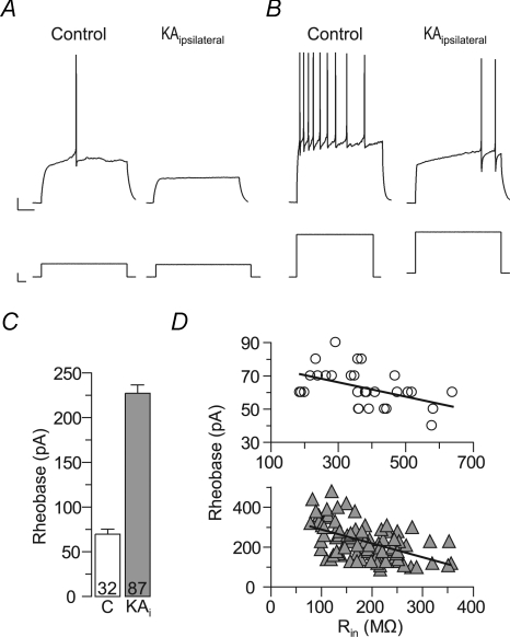
Figure 2. The excitability is reduced in epileptic granule cells of kainate-injected mice.
Epileptic granule cells of KA-injected (ipsilateral) hippocampi need more depolarizing current to generate action potentials (APs). A, a small current injection was sufficient to evoke an AP in control granule cells (left panel), but not in epileptic granule cells (right panel). B, the current needed to evoke minimal AP firing in epileptic granule cells (right panel) either killed control granule cells (not shown) or evoked strong AP firing in control cells (left panel). C, summary of experiments as in A and B. The current needed to generate at least one AP (rheobase) was strongly increased in epileptic granule cells (KAi). D, the rheobase was related to the Rin, suggesting that the latter was responsible for the reduced excitability of epileptic granule cells. Upper panel (circles), control granule cells; lower panel (grey triangles), epileptic granule cells). Note different y and x axis scales in these panels.

