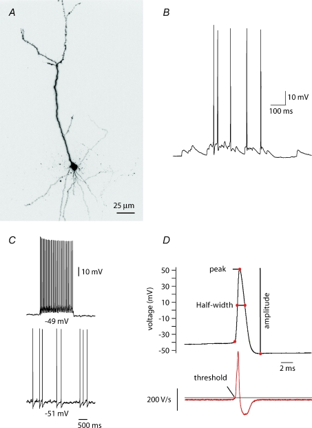
Figure 1. Cellular properties of presubicular cells.
A, 3-D reconstruction of the soma and principal dendrites of a Alexa Fluor 594-filled presubicular pyramidal cell. B, spontaneous EPSPs initiated action potentials. The membrane potential was −60 mV. C, depolarizing current injections to −51 mV (maintained) and −49 mV (step) initiated cell firing. D, action potential peak, half-width, amplitude and threshold measurements. The amplitude was defined as the spike height relative to the most negative voltage reached during the 20 ms following the peak. The half-width was taken at half-maximal spike amplitude. Threshold was defined as the voltage where dV/dt exceeds 10 V s−1.

