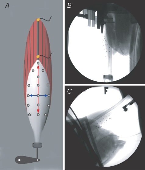
Figure 1. Methodological description of the in situ preparation used to quantify strain patterns in the lateral gastrocnemius aponeurosis of wild turkeys.
A, schematic diagram of the muscle preparation showing the location of the sonomicrometry crystals along a proximal muscle fascicle as well as the approximate position of the radio-opaque markers on the surface of the aponeurosis. The distal end of the tendon was attached to an ergometer, which controlled and measured muscle force and muscle–tendon length. B and C, single frames from videos captured using two high-speed fluoroscopes. The 3D positions of the surface markers were used to calculate the strain patterns in the aponeurosis during passive and active force production.

