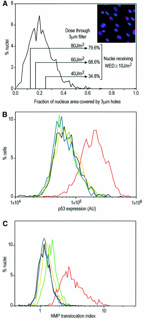
Fig. 2. Effects of micropore irradiation on p53 expression and NPM translocation in NDFs. (A) Distribution of the fraction of irradiated areas on NDF nuclei (n = 1699) observed by the ratio between the area exposed to UV irradiation through 3 µm Isopore filters (irradiated area detected by antibody labelling of photolesions) and the total nuclear projected area (Hoechst 33324). The insert shows an example field of micropore-irradiated nuclei, with CPDs labelled red and nuclei blue (Hoechst). (B) p53 expression levels in NDFs whole-nucleus irradiated at 10 J/m2 (red), micropore irradiated at 40 (green), 60 (yellow) and 80 J/m2 (blue), and non-irradiated (black). The fractions of nuclei receiving WED ≥10 J/m2 (see text) under each micropore irradiation condition are indicated in (A). (C) NPM translocation indext for NDFs irradiated in the same conditions as in (B). Cells in (A) were fixed immediately after irradiation, while cells in (B) and (C) were fixed 6 h after irradiation.

An official website of the United States government
Here's how you know
Official websites use .gov
A
.gov website belongs to an official
government organization in the United States.
Secure .gov websites use HTTPS
A lock (
) or https:// means you've safely
connected to the .gov website. Share sensitive
information only on official, secure websites.
