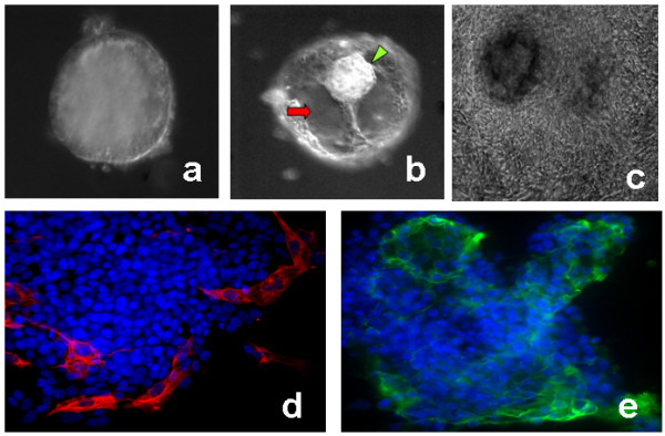
Figure 1.

Trophoblast differentiation from embryoid bodies on the biomimetic platform. Panel (a) shows a non-cystic early EB. Panel (b) shows a late cytic EB with clear fluid filled cavity (red arrow) and a small mass of cells similar to inner cell mass (ICM) of an embryos (green arrowhead) pushed towards the top. Panel (c) shows a cystic EB outgrowing on the biomimetic platform on day 16. Panels (d) and (e) immunolocalization of trophoblast markers cytokeratin 8/Troma1 (red) and SSEA1 (green) in cytic EB outgrowths at early day 8. Blue (DAPI stain) represent the nuclei. This shows the evidences for the first signs for clear and distinct trophoblast cell differentiation from cystic EBs as early as day 8.
