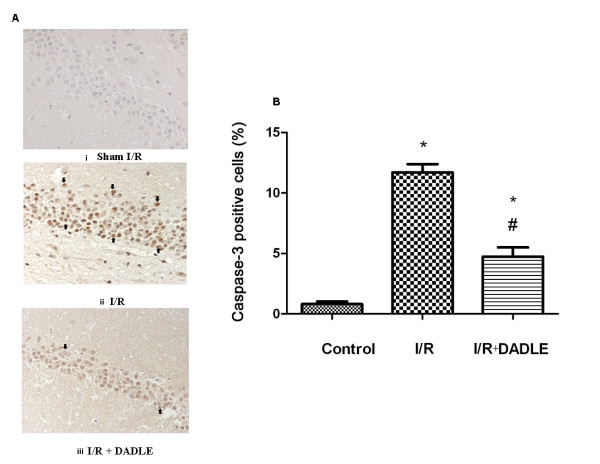
Figure 1.

[D-Ala2, D-Leu5]-Enkephalinamide (DADLE)-induced reduction of caspase 3 staining in the hippocampus exposed to ischemia/reperfusion (I/R) stress. A, Representative images. Strept actividin-biotin complex (SABC), 10 × 40. (i) Sham I/R. (ii) I/R. (iii) I/R plus. Arrows indicate caspase 3-positive cells. Note that there was little caspase 3 staining in the sham control slice, while there was strong signal staining in that of I/R group, which was largely reduced by δ-opioid receptor (DOR) activation with DADLE. B, Quantification of caspase 3-positive cells. Mean ± standard error (n = 6). *, P < 0.05, I/R or I/R + DADLE versus control. #, P < 0.05, I/R + DADLE versus I/R. Note that the administration of DADLE reduced the number of caspase 3-positive cells in the hippocampus following the cerebral ischemia/reperfusion.
