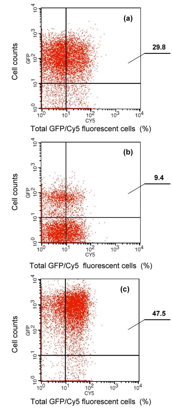
Figure 2.
Flow cytometry analysis of B. thuringiensis recombinant cells expressing (a) Mba-GFP, (b) Mbp-GFP and (c) Mbg-GFP fusion proteins. Cells were labelled with primary monoclonal anti-GFP antibody, followed by secondary Cy5-conjugated goat anti-mouse IgG. The histogram on the top right corner of each figure shows total GFP/Cy5-labelled fluorescent cells, and the values indicate their percentages of total cell counts. For each detection, a total of 100,000 cells were analyzed.

