Figure 3.
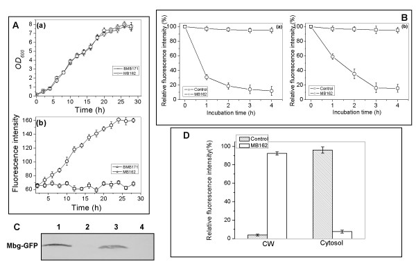
Expression profiles of B. thuringiensis MB162 cells. (A) Time course of (a) cell growth and (b) specific GFP fluorescence intensity of MB162 cells expressing Mbg-GFP fusion protein. Cells were cultured in LB at 30°C in the presence of 25 μg/mL of erythromycin, then harvested and diluted to unit cell density (OD600 = 1) with PBS buffer (pH7.0) for GFP fluorescence intensity determination. (B) SDS sensitivity and pronase accessibility assays of MB162 intact cells and the control cells expressing only cellular GFP. Relative values were based on GFP fluorescence intensity at the initial incubation time. Each value and error bar represents the mean of three independent experiments and its standard deviation. The values of MB162 cells without pronase or SDS treatment remained unvaried during the time course (data not shown). (C) Western blot analysis of MB162 cell fractions. Lane 1, cell-wall fraction; lane 2, soluble cytoplasmic fraction; lane 3, whole cell lysate; lane 4, BMB171 (the negative control). (D) GFP specific fluorescence intensity measurement of cell-wall (CW) and cell cytosol fractions.

