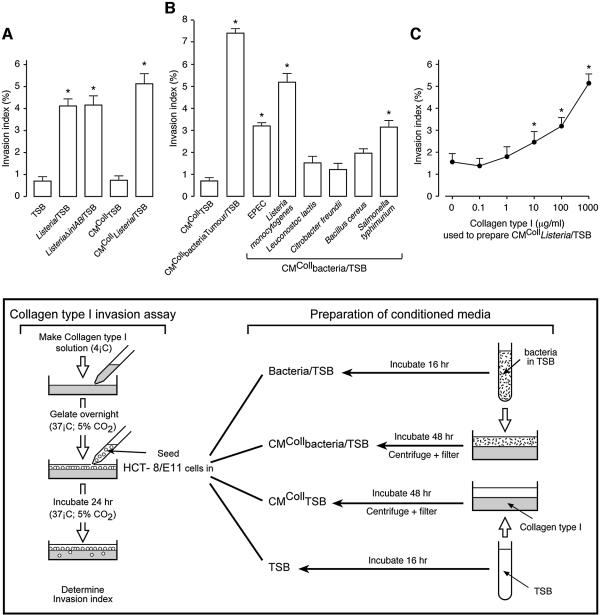
Fig. 1. Bacteria stimulate cancer cell invasion into collagen type I gels. (A) HCT-8/E11 cells were incubated, on top of collagen type I gels, in the presence of: TSB, L.monocytogenes wild-type or ΔinlAB mutant grown in TSB (Listeria/TSB or ListeriaΔinlAB/TSB, respectively), CM of TSB or of L.monocytogenes wild-type grown in TSB and produced for 48 h on top of collagen type I gels (CMCollTSB or CMCollListeria/TSB, respectively). The experimental design is presented schematically in the box. Invasion indices (%) were determined as in Materials and methods. Bars represent mean values of at least three independent experiments and flags indicate standard deviations. *Significantly different from TSB at p < 0.005. (B) HCT-8/E11 cells were incubated, on top of collagen type I gels, in the presence of CMColl of different bacterial strains grown in TSB and isolated from laboratory stock cultures or from the tumours of colon cancer patients (CMCollbacteria/TSB or CMCollbacteriaTumour/TSB, respectively). Invasion indices (%) were determined as in (A). *Significantly different from CMCollTSB at p < 0.005. (C) HCT-8/E11 cells were incubated, on top of collagen type I gels, in the presence of CMCollListeria/TSB, produced on gels with different collagen concentrations. Invasion indices (%) were determined as in (A). Dots represent mean values of at least three independent experiments and flags indicate standard deviations. *Significantly different from CMCollListeria/TSB prepared with 0 µg/ml of collagen type I at p < 0.005.

An official website of the United States government
Here's how you know
Official websites use .gov
A
.gov website belongs to an official
government organization in the United States.
Secure .gov websites use HTTPS
A lock (
) or https:// means you've safely
connected to the .gov website. Share sensitive
information only on official, secure websites.
