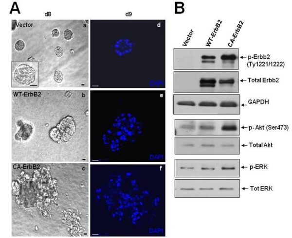
Figure 1.

Characterization of ErbB2 overexpression in normal MCF10A mammary epithelial cells. (A) Phase-contrast micrographs (a, b, c) or representative DAPI-stained equatorial confocal sections (d, e, f) of acinar structures formed from stable MCF10A cells infected with retrovirus expressing empty vector or the wild-type rat Neu/ErbB2 (Wt-ErbB2) or the constitutively active V664E Neu/ErbB2 mutant (CA-ErbB2) after 8-9 days of growth in Matrigel. Scale bars = 20 μm. (B) Western blots of whole-cell lysates from monolayer cultures of MCF10A cells expressing empty vector, Wt-ErbB2 or CA-ErbB2 constructs probed with the indicated antibodies to downstream targets of ErbB2 signaling. GAPDH was used as loading control.
