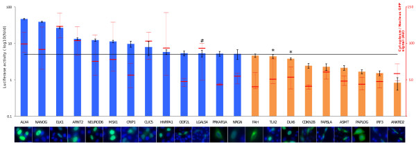
Figure 4.
Small-scale validation of the assay. Luciferase-based nuclear translocation assay and GFP-fusion nuclear localization assay were compared for 22 constructs. Histogram represents the log10 of the average luciferase ratio for three independent assays. Error bars are standard deviation. The black line represents the 5-fold threshold above which a given construct is qualified as able to translocate into the nucleus; histograms in blue highlight positive luciferase results and those in orange negative results. The (#) and (x) signs, respectively, highlight the false-positive and false-negative results when compared to GFP-fusion-based nuclear localization. A representative picture of the GFP-fusion assay with blue DAPI straining and green GFP is positioned under each tested construct. The red line and error bars represent the ratio of GFP intensity in the nucleus to that of the cytoplasm computed from the GFP-fusion-based nuclear localization images. Values are also summarized in Additional File 4.

