
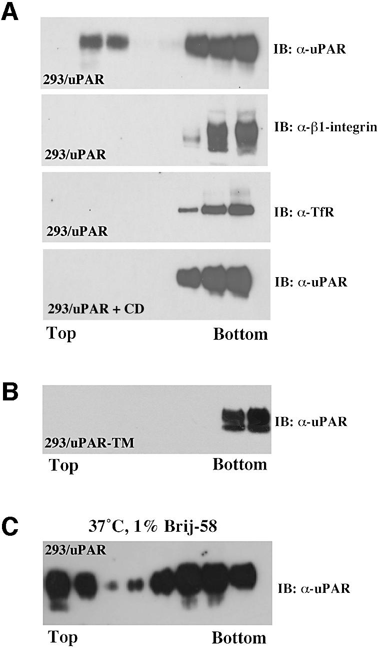
Fig. 1. uPAR partitions to two biochemically distinct membrane domains. (A) Western blot analysis of uPAR membrane localization. 293/uPAR cells were lysed in buffer containing 1% Triton X-100 and subjected to sucrose density gradient ultracentrifugation (see Materials and methods). Equal volumes of the resulting fractions (10 µl) were probed for uPAR using a polyclonal anti-uPAR antibody in immunoblotting. The same fractions were also probed with polyclonal antibodies to β1-integrin and the transferrin receptor, respectively. 293/uPAR cells were first treated with CD (10 mM, 1 h, 37°C) and then subjected to flotation analysis as described above. (B) 293/uPAR-TM-expressing cells were subjected to the same analysis. (C) 293/uPAR cells were detergent lysed in buffer containing 1% Brij-58 at 37°C and subjected to flotation analysis (see Materials and methods). The data presented are representative of several independent experiments.