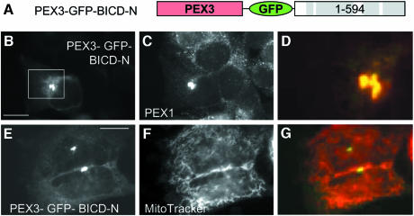
Fig. 2. BICD2 N–terminus, fused to PEX3, relocalizes peroxisomes, but not mitochondria. (A) Schematic representation of the PEX3–GFP–BICD-N fusion construct. (B–G) HeLa cells were transfected with PEX3–GFP–BICD-N fusion construct and stained for peroxisomes (C) or mitochondria (F). GFP signals are shown in the left panel, stained organelles in the middle panels, and an overlay (with GFP signal in green, organelles in red) in the right panel. Bar, 10 µm.

An official website of the United States government
Here's how you know
Official websites use .gov
A
.gov website belongs to an official
government organization in the United States.
Secure .gov websites use HTTPS
A lock (
) or https:// means you've safely
connected to the .gov website. Share sensitive
information only on official, secure websites.
