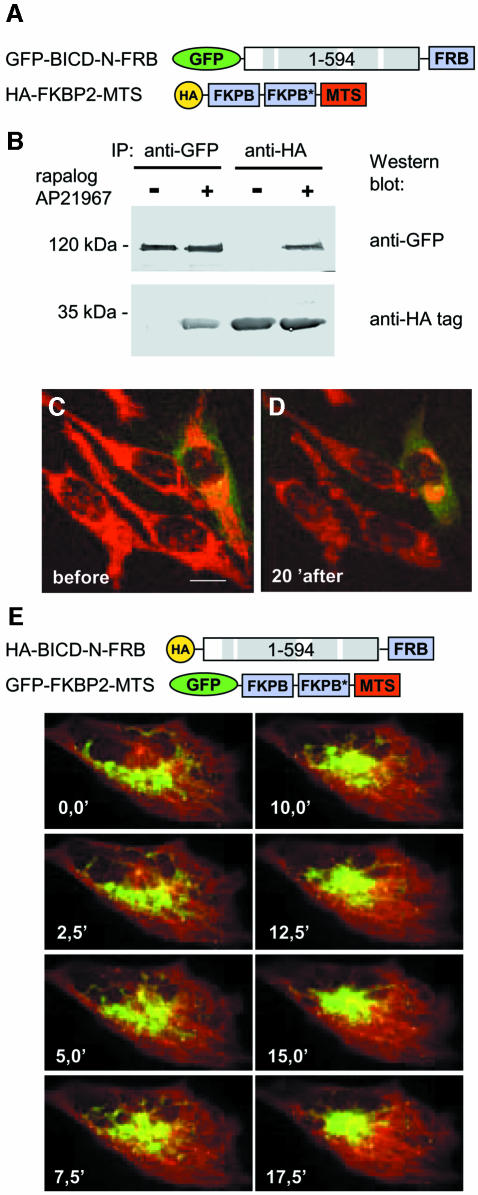
Fig. 3. Inducible targeting of BICD N–terminus to mitochondria, using regulated heterodimerization system. (A) Schematic representation of the fusion constructs. In the HA–FKBP2–MTS fusion the two FKBP domains differ by a number of synonymous substitutions, which reduce the recombination potential between the two tandem repeats (indicated by an asterisk). (B) HeLa cells were cotransfected with the two constructs, shown in (A), and used for immunoprecipitation with anti-GFP or anti-HA antibodies, and the immunoprecipitates were analyzed by western blotting. Where indicated with ‘+’, AP21967 (125 nM) was added to the cells 2 h before preparing the lysate. (C and D) HeLa cells, transfected with GFP-BICD-N-FRB and HA-FKBP2-MTS, were analyzed 1 day after transfection by confocal microscopy. Cells were stained for mitochondria with MitoTracker Red CMXRos for 30 min, washed and maintained in normal culture medium at 37°C. Cells before (C) and 20 min after the addition of 125 nM AP21967 (D) are shown. The transfected cell can be distinguished by green cytoplasmic signal, corresponding to the GFP–BICD-N–FRB fusion protein. (E) CAR fish fibroblasts were transfected with HA-BICD-N-FRB and GFP-FKBP2-MTS. Forty-eight hours after transfection, cells were microinjected with Cy3–tubulin. Cells were treated with 250 nM AP21967 and images were taken at the indicated times after the dimerizer addition. Note that the MTOC is clearly visible as the point of microtubule convergence. Bar, 10 µm.

An official website of the United States government
Here's how you know
Official websites use .gov
A
.gov website belongs to an official
government organization in the United States.
Secure .gov websites use HTTPS
A lock (
) or https:// means you've safely
connected to the .gov website. Share sensitive
information only on official, secure websites.
