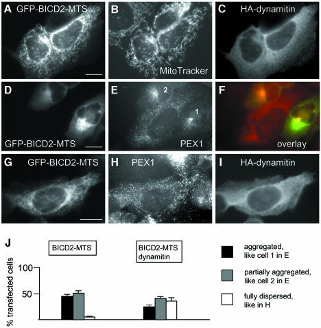
Fig. 7. Dynamitin inhibits aggregation of mitochondria and peroxisomes, caused by BICD2-MTS. (A–C) HeLa cells were cotransfected with GFP-BICD2-MTS and HA-dynamitin, stained with MitoTracker, fixed and stained for HA-tag. GFP signal is shown in (A), MitoTracker signal in (B) and HA-specific signal in (C). (D–F) HeLa cells were transfected with GFP-BICD2-MTS (D) and stained for peroxisomal marker PEX1 (E), overlay is shown in (F) (GFP signal in green, PEX1 signal in red). (G–I) HeLa cells were cotransfected with GFP-BICD2-MTS (G) and HA-dynamitin and stained for PEX1 (H) and HA-tag (I). Bar, 10 µm. (J) Quantitative analysis of the transfection results, shown in (D–I), performed as described for Figure 6P. Only cells which had a healthy, spread appearance and expressed medium levels (5–10 times compared with the endogenous BICD2 levels, as determined by counterstaining with anti-BICD2 antibodies), were scored. Staining for peroxisomes, rather than mitochondria, was used here for quantification, because it allows better assessment of the partially and fully dispersed phenotypes.

An official website of the United States government
Here's how you know
Official websites use .gov
A
.gov website belongs to an official
government organization in the United States.
Secure .gov websites use HTTPS
A lock (
) or https:// means you've safely
connected to the .gov website. Share sensitive
information only on official, secure websites.
