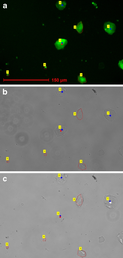
Fig. 2.
Laser pressure catapulting of detected male buccal cells. a Pseudo-colored image of the cell mixture: a blue catapulting dot is set on the male buccal cells, while the female cells and the debris are outlined in red. b Brightfield image after removal of the mounting medium and coverslip: male and female cells can easily be distinguished by the blue catapulting dots and the red outlining. c Brightfield image after LPC of the central male buccal cell

