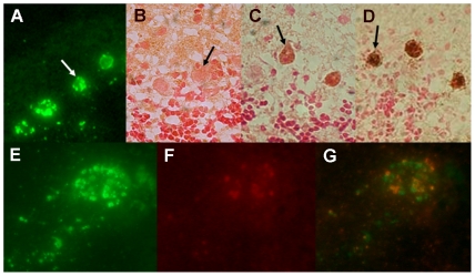
Figure 3. Rabies virus specific antibodies administered to CVS-F3-infected JHD−/− mice have limited access to infected CNS tissues.
At 7 and 9 days post-infection, CVS-F3-infected JHD −/− mice were treated with 1 mg of the rabies glycoprotein-specific, virus-neutralizing mouse monoclonal antibody 1112 (A, C, D, E, F, G) or saline vehicle alone (B). Several hours after the second treatment mice were transcardially perfused, CNS tissues removed and sections from the cerebellum were stained for rabies virus infection with FITC-anti-nucleoprotein monoclonal antibody (panels A and E; green), polyclonal rabbit anti-mouse biotinylated IgG (panels B and C; brown), additional 1112 antibody followed by the polyclonal rabbit anti-mouse biotinylated IgG (panel D; brown), or a combination of FITC-anti-nucleoprotein monoclonal antibody and rhodamine-conjugated anti-mouse Ig. Arrows in panels A-D identify Purkinje cells, a cell population that is extensively infected. Panels E-G show close-ups of a single cell stained with FITC-anti-nucleoprotein monoclonal antibody and rhodamine-conjugated anti-mouse Ig photographed using filters for FITC (panel E), rhodamine (panel F) and a combination red/green filter that shows both stains simultaneously (panel G).

