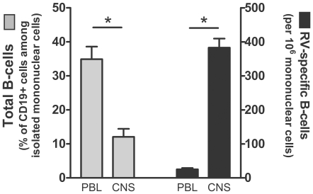
Figure 6. Rabies virus-specific antibody secreting B cells accumulate in the CVS-F3-infected CNS.
Mononuclear cells isolated from peripheral blood lymphocytes (PBL) and brain (CNS) were assessed for surface phenotype using flow cytometry and for the numbers of rabies virus-specific antibody secreting cells by ELISPOT. The percentage of B cells, identified as positive for both CD19 and MHC class II, among total mononuclear cells is presented on the left side of the figure while the proportion of the total mononuclear cells producing rabies virus-specific antibodies is presented on the right side of the figure.

