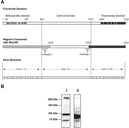
Figure 3.
POLQ domain structure, conservation and exon arrangement. (A) In the top line of the diagram, the position of a conserved helicase-like domain near the N-terminus of POLQ is shown with gray shading (black vertical bars show the positions of conserved motifs), and the position of a DNA polymerase domain is shown with black shading (white vertical bars show the positions of conserved motifs). Shaded regions in the middle line of the diagram show regions of the protein that are most highly conserved (see Fig. 6 for further detail). At the bottom, the exon structure of the protein is diagrammed. Exon 16 encodes residues 843–1879 of POLQ. Polyclonal antibodies 1 and 2 were raised against regions indicated by brackets. (B) HeLa S3 extract was analyzed by immunoblotting with polyclonal antibodies raised against the central domain of POLQ (positions indicated in the middle line in A). Lane 1, antibody 1; lane 2, antibody 2.

