Figure 3.
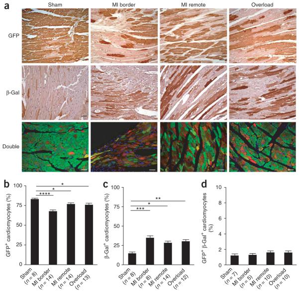
Stem or progenitor cells replenish adult mammalian cardiomyocytes after myocardial injury. (a) The percentage of GFP+ cardiomyocytes decreased after myocardial injury. MerCreMer-ZEG mice with 4-OH-tamoxifen pulse labeling received experimental myocardial infarction or left ventricular overload. After 3 months, mouse hearts were fixed and stained with antibodies to GFP (green) or to β-galactosidase (red) (double staining in yellow). Shown are representative images of GFP and β-galactosidase staining (brown) in hearts after sham operation, myocardial infarction (border and remote areas), and pressure overload. (b) Percentage of GFP+ cardiomyocytes. (c) Percentage of β-galactosidase+ cardiomyocytes. (d) Percentage of GFP+ β-galactosidase+ cardiomyocytes. *P < 0.05; **P < 0.01; ***P < 0.001; ****P < 0.0001. Scale bars, 20 μm. Data shown as mean ± s.e.m.

