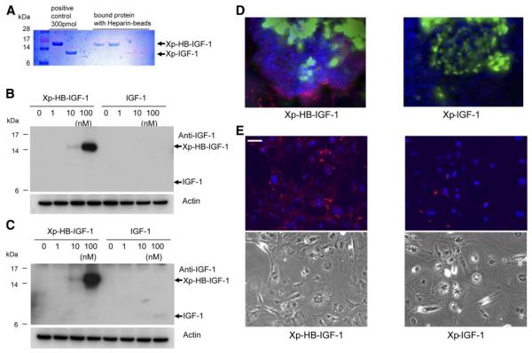
Figure 2.
Xp-HB-IGF-1 binds heparin and is retained more strongly in cell cultures. A) 300 pmol Xp-HB-IGF-1 or Xp-IGF-1 was incubated with heparin agarose beads for 2 h and washed 3× with PBS. Fusion proteins retained on the beads were detected by Coomasie blue staining. B, C) Doses of Xp-HB-IGF-1 or control IGF-1 were incubated with 3T3 fibroblast cells (B) or neonatal rat cardiac myocytes (C) for 2 h. The cells were collected after washing 3× with PBS, and the protein retained by the cell monolayer was detected by Western blot analysis using an anti-IGF-1 antibody. The results shown are representative of results from 3 independent experiments. D) Embryoid bodies were incubated with 100 nM Xp-HB-IGF-1 or Xp-IGF-1 for 2 h and then washed 3× with PBS. Retained protein was detected by immunofluorescent chemistry with an anti-Xpress antibody (red: anti-Xpress; green:α-myosin heavy chain; blue: DAPI). E) Embryoid bodies were dissociated by trypsin and EDTA, and monolayer differentiated ES cells were incubated with 100 nM Xp-HB-IGF-1 or Xp-IGF-1 for 2 h and then washed 3× with PBS. Retained protein was detected by immunofluorescence with an anti-Xpress antibody (red: anti-Xpress, blue: DAPI). Bottom panels show phase contrast. Scale bar = 50 μm.

