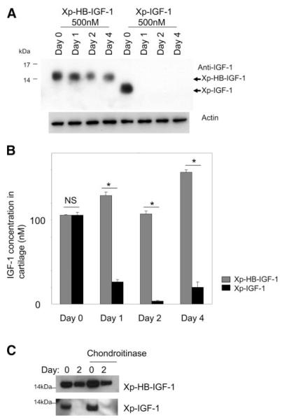
Figure 4.
Xp-HB-IGF-1 is retained in cartilage tissue. A, B) Cartilage disks were incubated with 500 nM Xp-HB-IGF-1 or Xp-IGF-1 for 2 days, then washed 3× with PBS and changed to media with no IGF-1 (day 0). IGF-1 fusion proteins remaining in the cartilage were detected by Western blot analysis with an anti-IGF-1 antibody (A) or by an ELISA using anti-Xpress and anti-IGF-1 antibodies (B). Results are expressed as mean ± se; n = 4. *P < 0.01. NS, not significant. C) No treatment cartilage disks or chondroitinase-treated cartilage disks were incubated with 500 nM Xp-HB-IGF-1 or Xp-IGF-1 for 2 days, then washed 3× with PBS and changed to media with no IGF-1 (day 0). IGF-1 fusion proteins remaining in the cartilage were detected by Western blot analysis with an anti-IGF-1 antibody.

