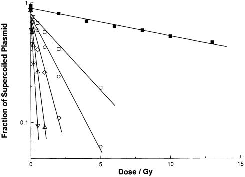
Figure 1.
Loss of supercoiled plasmid with increasing dose of γ-radiation. Aliquots of a solution containing plasmid pHAZE (25 µg ml–1), sodium phosphate (5 × 10⁻³ mol dm–3, pH 7.0), sodium thiocyanate (10–3 mol dm–3), sodium perchlorate (1.1 × 10⁻¹ mol dm–3) and tyrosine methyl ester [3 × 10⁻⁷ (open inverted triangle), 7.5 × 10⁻⁷ (open triangle), 1.5 × 10⁻⁶ (open diamond), 3 × 10⁻⁶ (open circle) or 5 × 10⁻⁶ (open and closed squares) mol dm–3] were irradiated under aerobic conditions with 137Ce γ-rays (662 keV) at dose rates between 1.7 × 10⁻³ and 9.0 × 10⁻² Gy s–1. After irradiation, the solutions were incubated for 30 min at 37°C with FPG at a concentration of 0 (closed square) or 5 µg ml–1 (open symbols). The fraction of supercoiled plasmid remaining after each radiation dose and incubation was determined using agarose gel electrophoresis. These six data sets are plotted together. Each is fitted to least mean squares straight lines of the form y = ce–mx. From the slopes m of these fitted straight lines, the doses D0 and SSB yields for the six irradiation and incubation conditions are: open inverted triangle, 0.234 Gy, 1.59 × 10⁻² µmol J–1; open triangle, 0.463 Gy, 8.04 × 10⁻³ µmol J–1; open diamond, 0.971 Gy, 3.84 × 10⁻³ µmol J–1; open circle, 1.94 Gy, 1.92 × 10⁻³ µmol J–1; open square, 3.41 Gy, 1.09 × 10⁻³ µmol J–1; closed square, 22.5 Gy, 1.66 × 10⁻⁴ µmol J–1.

