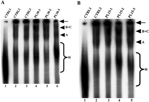
Figure 4.
Effect of the PLRG1 peptides on spliceosome assembly. Splicing reactions were performed for 50–60 min at 30°C and the splicing complexes formed were separated on a polyacrylamide/agarose native gel. The bands representing splicing complexes were revealed by autoradiography. (A) Lane 1, a control splicing reaction incubated on ice; lane 2 (CTRL2), a control splicing reaction without peptide; lane 3, the HC-2 peptide; lanes 4–6, the PL30-1, PL30-2 and PL30-3 peptides, respectively. (B) Lane 1, similar to CTRL1 in (A); lane 2 (CTRL2), the HC-2 peptide; lanes 3–5, the PL15-1, PL15-5 and PL15-5 peptides, respectively. The arrows on the right of the panels indicate material that did not penetrate into the gel. The arrowheads represent the splicing complexes separated on the native gel. The braces (labeled H) show non-specific complexes assembled on the pre-mRNA.

