Figure 7.

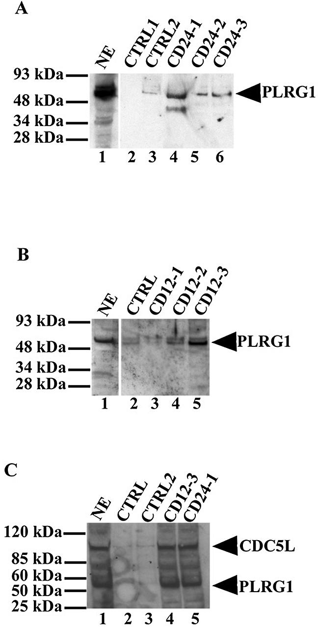
CDC5L peptides associate with PLRG1 in HeLa nuclear extract. CDC5L peptides were used in pull-down assays on streptavidin–agarose beads and the co-precipitated protein transferred to the nitrocellulose membranes was probed with anti-PLRG1 antibodies. Protein bands were then revealed by enhanced chemiluminescence (ECL). (A) Lane 1, a positive control containing HeLa nuclear extract; lanes 2 and 3, control pull-downs using beads only and CD-R24 peptide, respectively; lanes 4–6, protein from pull-down assays using the CD24-1, CD24-2 and CD24-3 peptides, respectively. (B) Pull-down assays using 12mer CDC5L peptides. Lane 1, a control containing HeLa nuclear extract; lane 2, a control pull-down assay using the CD-R12 peptide; lanes 3–5, protein from pull-down assays using the CD12-1, CD12-2 and CD12-3 peptides, respectively. (C) Binding of CDC5L peptides to PLRG1 in nuclear extract does not disrupt the CDC5L–PLRG1 interaction. Pull-down assays were performed as above using streptavidin–agarose beads except that the blots were probed with a buffer containing both anti-PLRG1 and anti-CDC5L antibodies. Lane 1, the positive control (HeLa nuclear extract); lanes 2 and 3, control pull-downs using the CD-R12 and CD-R24 peptides, respectively; lanes 4 and 5, pull-downs performed using the CD12-3 and CD24-1 peptides, respectively. The arrowheads on the right of the figure point to the bands representing PLRG1 or CDC5L on the nitrocellulose membrane.
