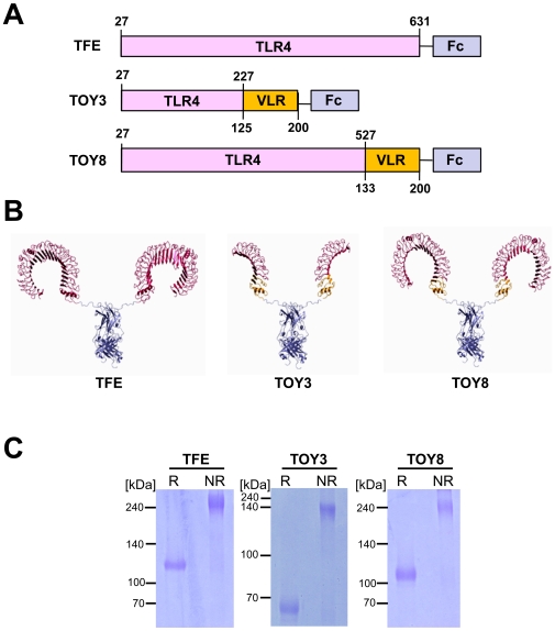
Figure 1. Structures of TFE and TOY constructs.
(A) Schematic diagrams of the constructs showing the relative sizes of the human TLR4 ectodomain (TLR4), VLRB.61 fragment (VLR) of hagfish, and human IgG-Fc (Fc). Numbers indicate amino acids of the parental proteins. (B) Crystal structures based on computer modeling. The domains depicted are TLR4 (red), VLR (yellow), and Fc (blue). (C) Each 2 µg of reduced (R) and nonreduced (NR) proteins was separated by SDS-PAGE and stained with Coomassie blue. Molecular masses (kDa) are indicated at left.

