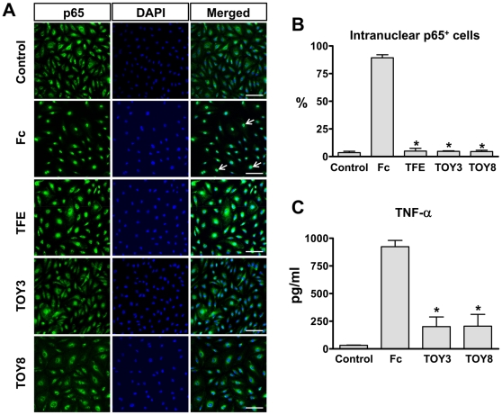
Figure 3. Pre-incubation of TFE, TOY3, and TOY8 markedly attenuates LPS-induced NF-κB activation in primary cultured lymphatic endothelial cells and TNF-α secretion in peritoneal macrophages.
LEC (primary lymphatic microvascular endothelial cells derived from human adult dermis) were purchased from Cambrex Inc. (East Rutherford, NJ) and maintained in endothelial cell basal medium-2 with growth supplements (EBM-2 MV). Passage 4–6 LEC were incubated in EBM-2 MV containing 1 % FBS for 8 hr and then with 1 µg/ml of Fc, TFE, TOY3, or TOY8 for 15 min, and then the LEC were treated with LPS (500 ng/ml) for 30 min. (A) For determination of NF-κB activation, nuclear translocalization of p65 (a subunit of NF-κB) was analyzed by immunostaining (green). Nuclei were counterstained with DAPI (blue). Arrows indicate nuclear translocalization of p65. Scale bars, 100 µm. (B) Cells positive for p65 intranuclear staining (white arrows) were counted among 100 cells arbitrarily chosen in 4 different regions, and the values presented as a percentage of the total cell number. Bars represent means ± S.D. (n = 4). *, P<0.05 versus Fc. (C) Primary cultured macrophages from mouse peritoneal cavity were pre-treated with 1.0 µg/ml of Fc, TOY3, or TOY8 for 30 min, and then were treated with LPS (100 ng/ml) for 4 hr. Culture media were sampled, and levels of TNF-α were measured. Bars represent means ± S.D. (n = 5). *, P<0.05 versus Fc.

