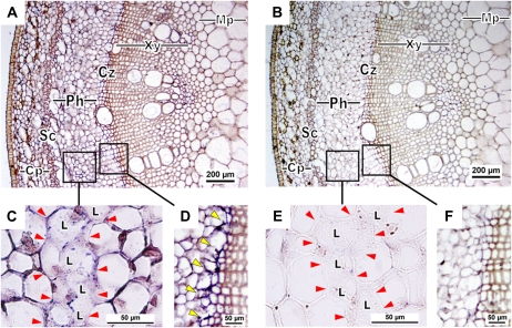
Figure 8.
In situ hybridization of HbPIP2;1 transcripts in young rubber stem. Transversal sections were hybridized with HbPIP2;1 antisense (A, C, and D) or sense (B, E, and F) digoxigenin probes. Transcripts of HbPIP2;1 are visualized by the intensity of blue to purple color developed by the reaction with anti-digoxigenin antibodies coupled to alkaline phosphatase using nitroblue tetrazolium/5-bromo-4-chloro-3-indolyl phosphate as substrates. The different images correspond to increasing magnifications in the same area. Sense probe control was used to indicate unspecific background signaling. Cp, Cortical parenchyma; Cz, cambium zone; L, laticiferous cell; Mp, medullar parenchyma; Ph, phloem; Sc, sclerenchyma; Xy, xylem vessel. The yellow arrowheads indicate the labeled precambial area in D. The red arrowheads indicate the shape of the fusing young latex cells (laticifers) in C and E. The probe concentration was 3 μg mL−1. Bars = 200 μm (A and B) and 50 μm (C–F).

