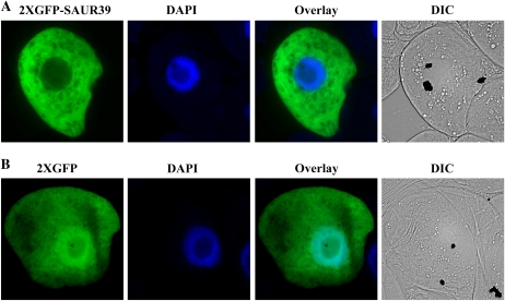
Figure 5.
Subcellular localization of transiently expressed 2XGFP-SAUR39 (A) and 2XGFP (B) proteins. The plasmids were transformed biolistically into tobacco BY2 cells, and images were observed by epifluorescence microscopy. From left to right are the subcellular localization of 2XGFP-SAUR39 or 2XGFP fusion protein, nuclei stained with 4′,6-diamidino-2-phenylindole (DAPI), overlay of the two images, and differential interference contrast (DIC) images.

