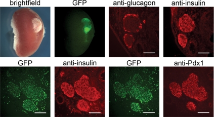
Figure 1.
Mature islets develop from transplanted fetal pancreatic anlagen. E12.5 fetal pancreatic anlagen transplanted under the kidney capsule of a prediabetic NOD mouse and recovered after 20 d develop into mature islets. The transplanted tissue is evident under the kidney capsule as shown in the bright-field and GFP fluorescence images at the upper left (not all of the anlagen transplanted were GFP-positive). Adjacent frozen sections of the graft site stained with antiglucagon and antiinsulin antibodies show insulin-positive β-cells surrounded by glucagon-positive α-cells in mature islets, shown in the right two images in the upper panel. Adjacent frozen sections showing GFP fluorescence and either insulin or Pdx1 expression are in the lower panels. The GFP marks cells of graft origin, whereas the localization of insulin and Pdx1 indicate that a subset of the GFP-positive cells have differentiated as β-cells. Bar, 100 μm.

