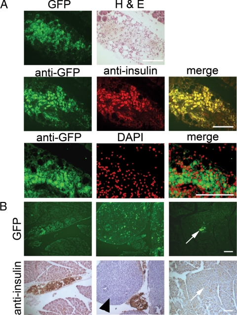
Figure 4.
Embryonic pancreatic precursor-derived cells colonize the host pancreas and form insulin-producing β-cells. A, Frozen sections through the pancreas of a diabetic NOD mouse that received the combination treatment and was unilaterally nephrectomized at 45 d. Top row, GFP fluorescence and H&E staining on the same section. Exocrine tissue is evident in the top corners. Middle row, Colocalization of GFP and insulin as evidenced by two-color immunohistochemistry with a merged image. Bottom row, Dual staining with anti-GFP and 4′,6-diamidine-2-phenylindole dihydrochloride (DAPI; false colored) with a merged image. Bar, 100 μm. B, GFP fluorescence (upper panels) and immunohistochemical analysis with antiinsulin (lower panels) was carried out on frozen sections of pancreas from diabetic NOD mice that received the combination treatment and were unilaterally nephrectomized at 45 d. Panels show several areas with different morphology of GFP-positive, insulin-positive clumps of cells in the interlobular spaces or attached to the endogenous pancreas. Additionally, the middle and right pairs of panels show examples of GFP-positive, insulin-negative cells in a large clump in the interlobular space (arrowhead) and a small clump integrated within the exocrine pancreas (arrows). Bar, 100 μm.

