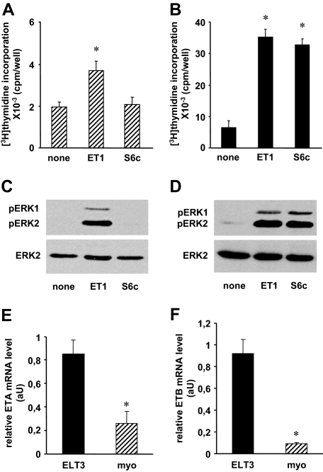
Figure 1.
Differential expression and function of ETA and ETB in myometrial and ELT3 cells. A and B, [3H]thymidine incorporation. Myometrial (A) and ELT3 (B) cells were treated or not for 48 h with or without 50 nm ET-1 or 50 nm S6c. [3H]thymidine (2 μCi/ml) was added 24 h before the end of the incubation, and [3H]thymidine incorporation was measured as described in Materials and Methods. Values are the mean ± sem of four (myometrial cells) or six (ELT3 cells) separate experiments performed in duplicate. *, P < 0.05 vs. untreated cells. C and D, ERK1/2 activation. Myometrial (C) and ELT3 (D) cells were incubated at 37 C for 5 min with or without 10 nm ET-1 or 10 nm S6c. Cells were lysed and detergent-extracted proteins were analyzed by 10% SDS-PAGE followed by immunoblotting with antiactive ERK1/2 antibody. After stripping, the blots were reprobed with antitotal ERK2 antibody (ERK2). Result of a typical experiment is among three performed. E and F, ETA (E) and ETB (F) mRNAs in myometrial (myo) and ELT3 cells were quantified by quantitative RT-PCR as described in Materials and Methods. Values, expressed as relative mRNA amounts normalized to total RNA, are the mean ± sem of three separate experiments performed in duplicate. *, P < 0.05 vs. ELT3 cells. aU, Arbitrary units; pERK1, phosphorylated ERK1.

