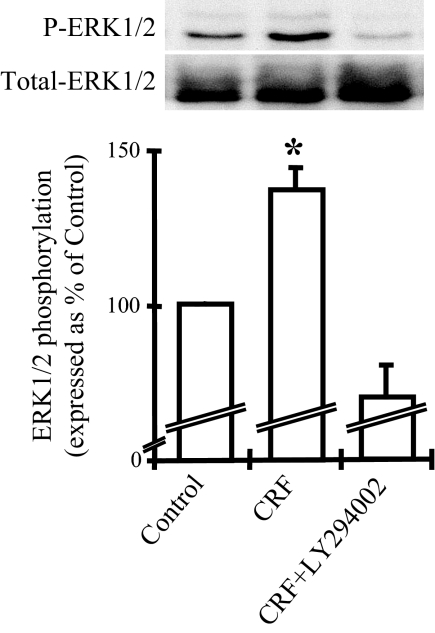
Figure 3.
Inhibition of the PI3K/Akt pathway abolishes the CRF-induced ERK1/2 phosphorylation. THP-1 cells were stimulated with CRF (10−7 m) alone or costimulated with LY294002 (30 μm) for 5 min. ERK1/2 phosphorylation was examined by Western blot analysis. A representative Western blot of the phosphorylated (P-ERK1/2) and total ERK1/2 is presented on the top together with a graphical representation of the combined data of four individual experiments. Phosphorylated ERK1/2 expression was standardized to total ERK1/2 and represented as percentage of the control. *, P < 0.05 relative to control.

