
Figure 2.
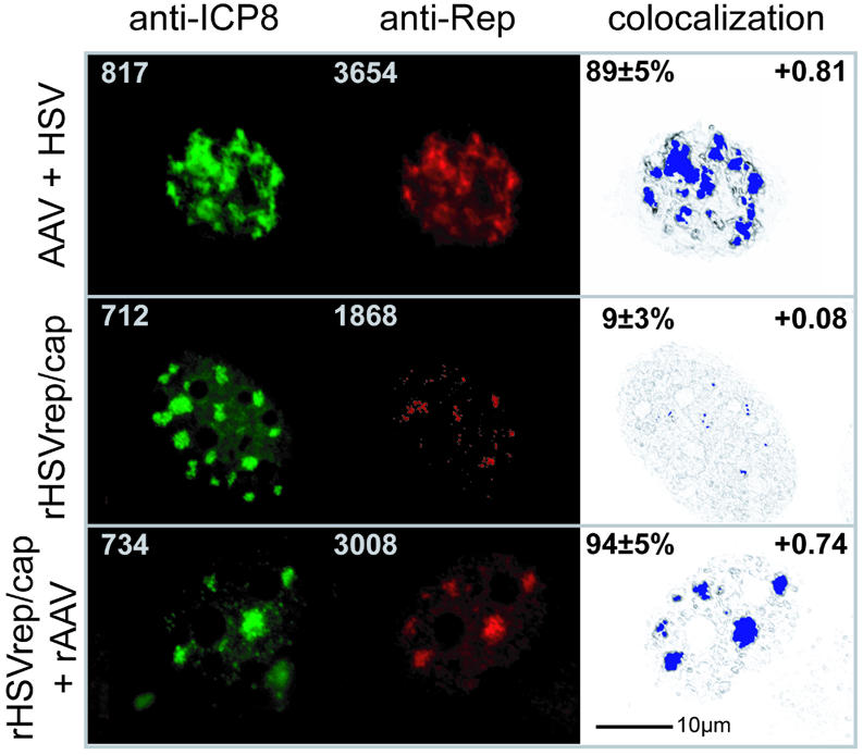
Subnuclear colocalization of AAV Rep and HSV ICP8. BHK cells were grown on coverslips, infected as indicated and fixed at 5 h p.i. Anti-Rep hybridoma 76.3 reactivity was visualized with Cy3-conjugated F(ab)-fragments of goat anti mouse IgG in the ‘red’ channel. The anti-ICP8 hybridoma 39S was applied and detected with Alexa 350-conjugated goat anti-mouse IgG in the ‘blue’ channel. Since blue color poorly reproduces on printed color panels, the Alexa 350 staining is represented as green false color (anti-ICP8 panel). For 3D-colocalization analysis the raw data set was restored using the ICTM algorithm of the Huygens 2 software (see Materials and Methods). The digitally deconvolved 2-channel 3D image was exported to the colocalization 1 software and to ImarisColoc for quantitative analysis, respectively. Following channel definition, a 2D histogram was calculated and a polygonal region was selected. This region was chosen by creating a selection proposal from the AAV + HSV data set. The threshold values and the histogram region were stored and applied to all data sets. After calculating the colocalization map, the colocalization voxels were displayed in blue color. The standard deviation of the intensities of all colocalization voxels in a map was found to be ≤8%. To facilitate navigation within the image the highlighted colocalization voxels were merged to a summed image (colocalization panel). Values given in the upper left edges of green and red panels are maximum fluorescence intensities measured within the raw 3D data set. The percent of green channel volume colocalized with red was calculated in ImarisColoc (Bitplane AG) for n = 6 nuclei and is given as mean ± SD in the upper left edges of the colocalization panel. The Pearson channel correlation in colocalized volume is given in the upper right edges of the colocalization panel (1 is perfect colocalization, 0 no correlation and –1 perfect inverse correlation). Recombinant AAV is transfected pTR-UF5 DNA.