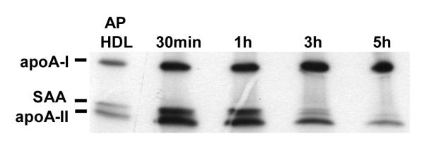
Figure 1B.
Preferential loss of apoA-II and SAA after SR-BI processing.
ApoA-I-/- mice were injected with 1.5×10¹¹ particles of adenoviral vector AdSR-BI. After three days when SR-BI expression was maximal, the mice were injected with a bolus of 750 μg 125I-AP HDL (25μg LPS). Plasma samples containing remnants were collected from individual mice at the indicated times after bolus injection and 5 μl aliquots were analyzed by SDS-PAGE (5-20% acrylamide gradient). An aliquot of the injected 125I-AP HDL is shown for comparison. Samples were visualized by autoradiography.

