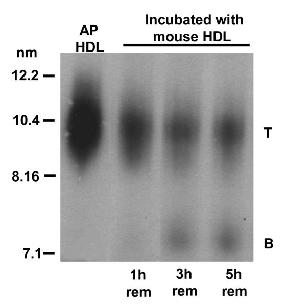
Figure 3A.
Size change of mouse AP HDL remnants that remodel by associating with mouse HDL.
Plasma samples containing HDL remnants, collected from a SR-BI-over expressing mouse 1, 3 and 5 hr after bolus 125I-AP HDL injection were mixed 1:10 (v/v) with C57BL/6 mouse HDL (1.7mg /ml) for 2 hr at 37° C to allow for remnant association with HDL. The resultant products were subjected to non-denaturing gel electrophoresis using a 4-18% acrylamide gradient and visualized by autoradiography. Mouse AP HDL is shown for comparison. T: remodeled remnant particles and B: remodeling-resistant particles.

