Figure 5.
Immunofluorescent staining of E14 control (A & B) and CatnbEx3(cko/+) (C & D) main-stem bronchi (MSB) with antibodies against β-catenin (C terminal, green) and β-catenin (N terminal, “wild type” red). Arrows indicate the polyps.
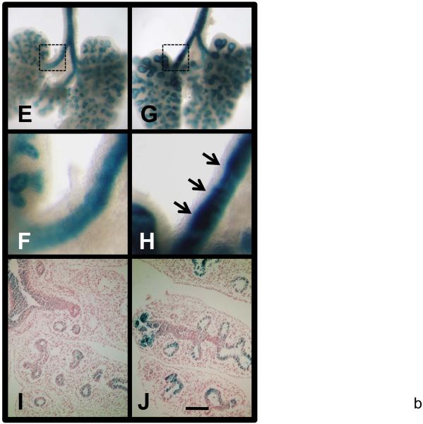
LacZ staining of E13 TOPGAL (E & F) and Nkx2.1-cre;Catnb[+/lox(ex3)];TOPGAL (G & H) lungs from siblings. F & H are higher magnification of boxed areas of E & G, respectively. I & J are sections of stained TOPGAL and Nkx2.1-cre;Catnb[+/lox(ex3)];TOPGAL lungs with eosin counter staining, respectively. Scale bar: 40 um (A-D); 1 mm (E, G); 250 um (F, H); 120 um (I, J).


