Figure 1.
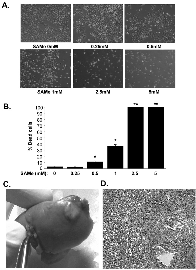
A and B) SAMe causes dose-dependent cell death in H4IIE cells. Cells were treated with varying doses of SAMe for 24 hours and phase contrast images are shown in A. while the graph in B. summarizes the results of 4 experiments shown as % of dead cells in mean±SEM. *p<0.001, **p<0.00001 versus (vs.) control (no SAMe). C) In vivo HCC model. Two weeks after injecting 2.5×10⁶ H4IIE cells into the liver parenchyma, a clearly visible tumor can be seen. D) HCC histology on H&E (100X) shows the tumor is composed of cells having round to oval nuclei, small nucleoli, basophilic cytoplasm, and increase in the nuclear:cytoplasmic ratio. The cells predominantly form sheets as well as in areas thickened cords in part lined by flattened endothelial cells. Occasional variably-sized vascular spaces are also seen. The adjacent non-tumor liver shows variable sinusoidal dilatation and congestion.

