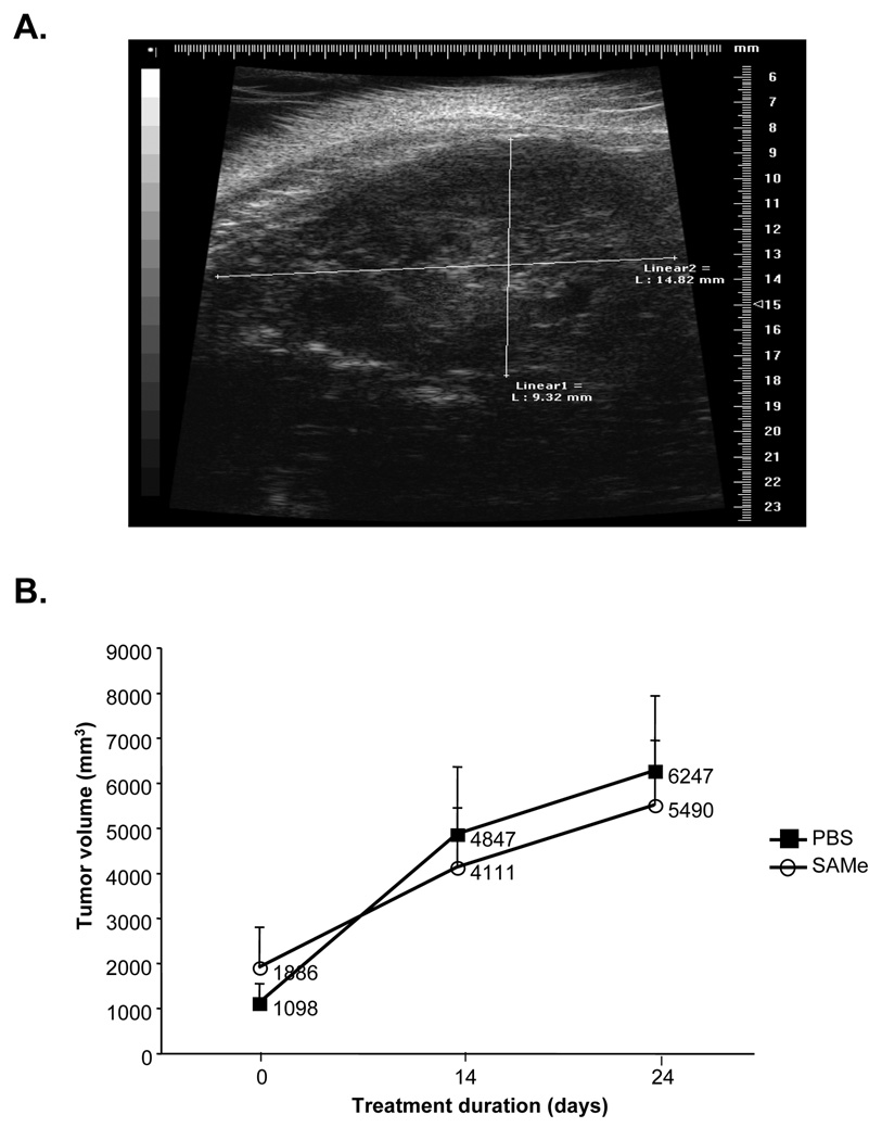
Figure 3.
A) Representative ultrasound image of a rat with HCC. B) Effect of IV SAMe treatment in already established HCC. Rats were previously injected with H4IIE cells (2.5×10⁶ cells) and imaged by ultrasound (one representative slice through the tumor). They were then divided into two groups (n=5 per group) with comparable starting tumor volumes and treated with IV SAMe (150mg/kg/day) or PBS for 24 days. Ultrasound imaging was performed in all rats on day 14 of treatment and on day 24, at which time all rats were sacrificed. Graph summarizes change in tumor volume with time.

