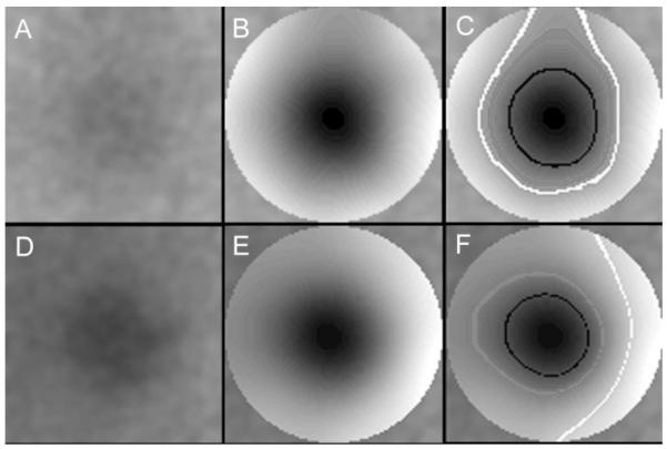
Figure 2.
Filtered foveal images and isobar patterns. (A) The foveal region of interest in Figure 1 was filtered on a small scale (Gaussian blur, radius 36 μm) for noise reduction. The central fovea was, on average, more hypofluorescent, but no pattern was apparent. (B) The fovea (1500-μm diameter disc) was further filtered (Gaussian blur, radius 180 μm) to establish a very regular shading pattern of concentric elliptical isobars of isofluorescence. Contrast enhancement was applied, to emphasize the geometry of the pattern. The individual isobars were still too fine to be discernible, and so three are highlighted (C) with black (original GL = 112, before contrast enhancement), gray (original GL = 120), and white (original GL = 123). There were 30 isobars in this pattern, yielding an average isobar resolution of 750 μm/30 = 25 μm. The isobars illustrated were even finer, mostly one pixel (15 μm) in width. In this typical pattern, the central isobars were nearly circular, with the more peripheral isobars becoming more vertically elongated until the last were only partial annuli. (D) Another normal foveal AF image of the left eye of a 53-year old woman after the small-scale filter was applied. The isobar pattern in (E) showed the temporal fovea to be slightly more hyperfluorescent than the other quadrants and the hypofluorescent center appeared somewhat elongated horizontally. These features were dramatically more evident in the individual isobars in (F), where isobars are highlighted in black (GL = 62), gray (GL = 75), and white (GL = 97). The ellipses in this less typical pattern became more elongated horizontally and finally became more hyperfluorescent arcs temporally. There were 36 isobars in all in this pattern, yielding an average isobar resolution of 21 μm. The isobars shown are 1 pixel (15 μm) in width, with occasional single pixel discontinuities.

