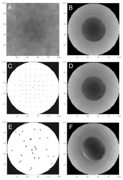
Figure 4.
Modeling and reconstruction of normal AF foveal background. The vertical and horizontal scales on all panels are in pixels. (A) Preprocessed foveal AF image of the left eye of a 50-year-old woman. (B) Contour graph of the model fit to the complete GL pixel data from the image in (A). (The software used for the analysis added the fine black lines to delineate individual contours, but they are not part of the model.) The coefficients (a, b, c, d, e, constant) of the inner and outer quadratics of the model were (0.0297, 0.0087, 0.0367, 0.0278517, -0.00725, 93.6) and (0.00692, -0.0006, 0.0108, -0.0709, 0.0381, 111.7), respectively. (C) Uniform grid of pixel data from (A)to which the model was fit independently. (D) Reconstruction of the foveal AF data from the grid subset of the pixel data in (C) by the model. The coefficients of the quadratics fit to the grid data were (0.0318, 0.00778, 0.0353, 0.0762, -0.0303, 93.5) and (0.00676, -0.0014, 0.0111, -0.0553, 0.0331, 111.6), respectively. The model was nearly identical with the model in (B). The mean absolute error of the model (D) with respect to the image in (A) was also nearly as good as that of (B; 3.7% compared with 3.6%). (E) Random subset of pixel data from (A) to which the model was fit independently. (F) Reconstruction of the foveal AF data from the random subset of pixel data in (E) by the model. The coefficients were (0.0302, 0.0441, 0.0450, -0.160, -0.271, 94.7) and (0.00725, 0.00084, 0.0129, -0.0753, 0.0843, 109.5). The resulting model was quite similar to the model in (B) except at the boundary transition inferonasally. The mean absolute error of the model (F) with respect to the image in (A) is somewhat increased compared with that of (B) (5.7% compared to 3.6%).

