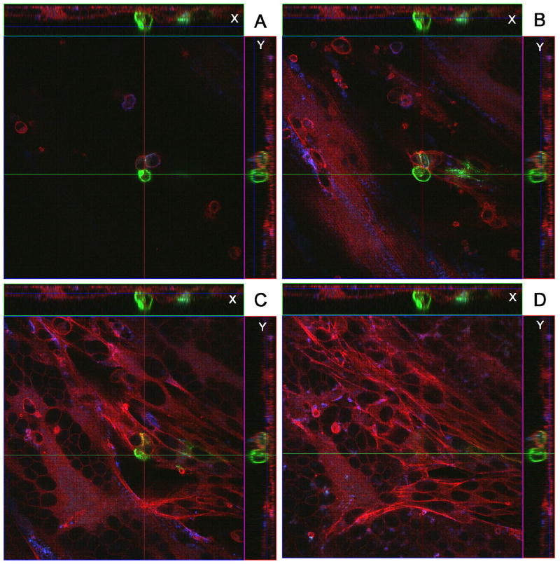
Fig. 5.
Identification of a VZV gC-positive inoculum cell at 48 hpi. All conditions were the same as described in legend to Fig. 4. The sample was then viewed in a Zeiss LSM 510 confocal microscope and a Z-stack (15 slices, 2 um thick) of images at 400 X was taken, showing a gC-positive inoculum cell on the cellular monolayer. Panels A–D show four slices from the Z-stack. Panel A is slice 11 (top most), B is slice 7, C is slice 5 and D is slice 3 (bottom most). Each panel includes the view from the Z direction with smaller side panels showing the view from X and Y, respectively. Note the small syncytium in the lower left corner of panels C and D, beneath plane of the gC-positive cell.

