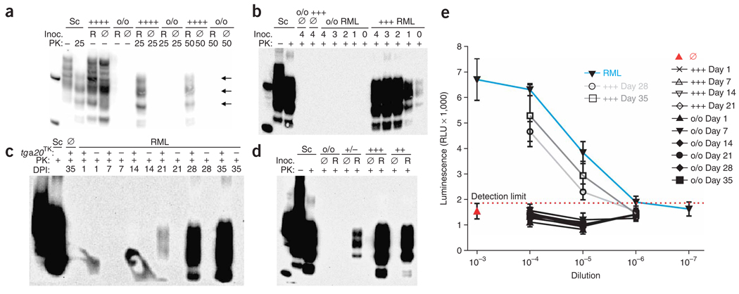
Figure 1.
Prions in slice cultures. (a) Immunoblot from tga20TK and Prnpo/o slices exposed to RML-infected (R) or uninfected (Ø) brain homogenate, cultured for 35 d, optionally digested with PK, and probed with antibody POM1 to PrP. Here and in all following figures, sample labels indicate the PrPC expression dictated by the slice genotype: o/o, Prnpo/o; +/−, hemizygous Prnp+/o; +/+, wild type; ++, hemizygous tga20 and Prnpo/o; +++, hemizygous for the tga20 allele and Prnp+/o; ++++, homozygous tga20 and Prnpo/o; Sc, positive-control homogenate from brain of a mouse with scrapie; inoc., inoculum. Left lane on all blots, molecular weight marker spiked with recombinant PrPC, yielding a PrP signal at 23 kDa with a cleavage product at 15 kDa. Arrows, PrPSc glycoforms. (b) Sensitivity of the POSCA. Immunoblot from tga20TK and Prnpo/o slices inoculated with decadal dilutions (104–100 µg; lane label, log(µg)) of prion-infected (RML) or uninfected (Ø) brain homogenate and cultured for 35 d before harvesting. (c) Time course of PrPSc accumulation in infected slices. Immunoblot from tga20TK (+) and Prnpo/o (−) slices inoculated with 100 µg prion-infected (RML) or uninfected (Ø) brain homogenate. (d) Immunoblot from Tga20TK, tga20+, Prnp+/o or Prnpo/o slices inoculated with 1 mg brain homogenate and harvested 35 dpi. (e) Time-dependent buildup of prion titers in slices. RML-infected or uninfected brain homogenate and slice homogenates from the experiment reported in panel c were serially tenfold diluted in uninfected brain homogenate, and infectivity was determined by transfer to PK1 cells. Means of three independent biological replicas ± s.d. RLU, relative light units.

