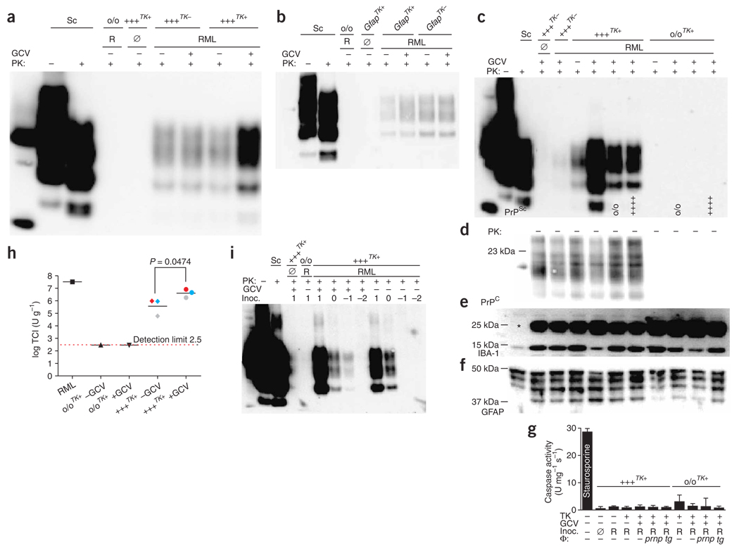
Figure 5.
Impact of microglial depletion on prion replication. Cultures were prepared from 10-d-old mice either CD11b-HSVTK+ (tga20TK+ or Prnpo/o/TK+) or CD11b-HSVTK− (tga20TK−). (a) Western blot from infected tga20TK− and tga20TK+ slices treated with GCV. Samples were PK-digested and PrP detected with POM1. (b) Western blot from cultures from 10-d-old GFAP-HSVTK− (GfapTK−), GFAP-HSVTK+ (GfapTK+) or Prnpo/o mice treated as in a. (c–h) Macrophage reconstitution of microglia-depleted tissue. Antigen detected is indicated in lower left corner of immunoblots. (c) Experiment performed as in a, showing marked increase in PrPSc accumulation upon GCV treatment of RML-infected tga20TK+ slices, which was counteracted by Prnpo/o or tga20+/+ macrophages (genotype at bottom of lanes). (d–f) Undigested sample detected with POM1 (d), rabbit anti-Iba1 (e; *unidentified cross-reacting protein) or mouse anti-GFAP (f). (g) Caspase-3 enzymatic activity, normalized to protein content. Averages of four biological replicas ± s.d. (NS, P > 0.05). (h) SCEPA of homogenates of tga20TK+ or Prnpo/o/TK+ slices treated as in a. Three independent biological replicas of tga20TK+ and single replicas of Prnpo/o/TK+ slices or RML were analyzed in tenfold dilution steps using 6–12 replica PK1-containing wells per dilution. Data are indicated as the number of infectious tissue culture units per gram of slice culture protein and are the averages of biological replicas ± s.d. Slices from the same animal (pairs of −GCV and +GCV samples) are represented by the same color. (i) Immunoblot from cultures from 10-d-old tga20TK+ or Prnpo/o/TK+ mice uninfected (Ø) or infected with RML in tenfold dilution steps from 1.7 to −1.3 log SCI50; log(µg) inoculum is indicated above each lane. Each sample was divided into two pools (−GCV and +GCV) and harvested 35 dpi.

