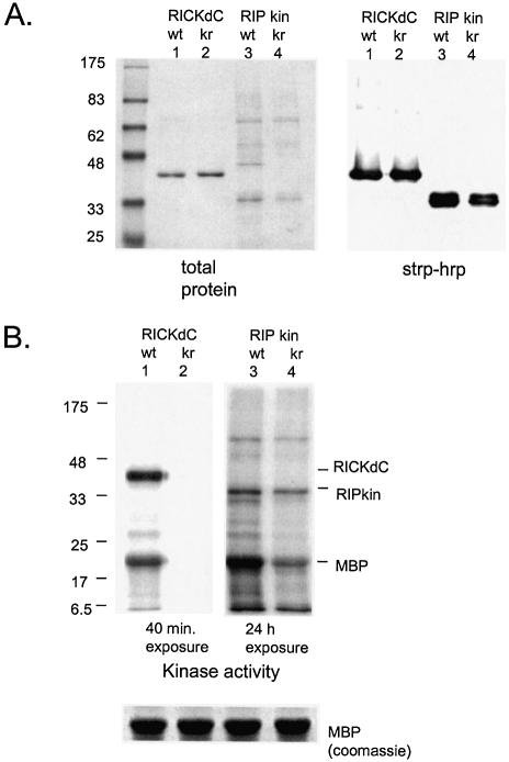
Figure 3.
Analysis of contaminating kinase activity. (A) Protein content. RICKdC and RIPkin (both wild type and kinase inactive forms) were expressed in 293 cells, extracted and purified by streptavidin–Sepharose. Similar quantities of each protein were resolved by SDS–PAGE and stained for total protein (left side) or transferred to nitrocellulose, stained with streptavidin–HRP followed by ECL (right side). (B) Kinase activity. Equal quantities of RICKdC (wt or K47R) and RIP kin (wt or K45R) were incubated under RIP kinase conditions (see Materials and Methods) in the presence of [γ-33P]ATP and MBP as a substrate. The protein reactions were resolved by SDS–PAGE, and the radioactive pattern was revealed by exposure to X-ray film for either 40 min (RICK samples) or 24 h (RIP samples). The bottom panel shows equivalent MBP staining by Coomassie blue in four parallel reactions.

