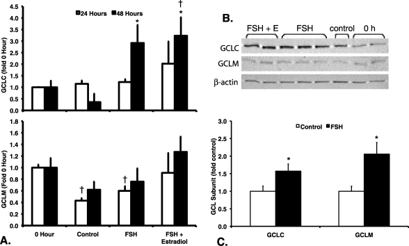
FIG. 2.
Effects of FSH and estradiol on GCL subunit protein levels in granulosa cells and small antral follicles. Granulosa cells and follicles were cultured as described for Figure 1, except that only the 20-ng/ml concentration of FSH was used. GCLC, GCLM, and β-actin protein levels were measured by immunoblotting of protein extracts from granulosa cells (A and B) and antral follicles (C) as described in Materials and Methods. Relative protein levels normalized to β-actin were expressed as fold of 0-h controls (A) or untreated controls (C) for the same blot. Data are expressed as means ± SEM. A) GCLC protein levels (upper graph) varied significantly by treatment at 48 h (P = 0.019 by Kruskal-Wallis test) but not at 24 h (P = 0.331 by Kruskal Wallis test). GCLM protein levels (lower graph) varied significantly by treatment at 24 h (P = 0.016 by Kruskal-Wallis test) but not at 48 h (P = 0.248 by Kruskal-Wallis test). †Significantly different from 0-h controls at the same time point, P < 0.05 by Mann-Whitney test. *Significantly different from untreated controls at the same time point, P < 0.05 by Mann-Whitney test (n = 3–5 per treatment group and time point). B) Representative Western blot showing GCLC, GCLM, and β-actin levels in granulosa cells after 48 h of culture with the indicated treatments. C) *Treatment of antral follicles with FSH for 24 h resulted in statistically significant increases in protein levels of GCLC (P = 0.045 by t-test) and GCLM (P = 0.022) compared with untreated controls (n = 6 observations of eight follicles each per treatment group).

