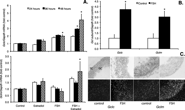
FIG. 3.
Effects of FSH and estradiol on GCL subunit mRNA levels in granulosa cells and small antral follicles. Granulosa cells and follicles were cultured as described for Figures 1 and 2 and were harvested at the indicated times for analysis of Gclc, Gclm, and Gapdh mRNA by quantitative real-time RT-PCR as described in Materials and Methods. The graphs show mean ± SEM levels of Gclc and Gclm normalized to Gapdh and expressed as fold change from the untreated control levels. A) When each time point was analyzed separately by Kruskal-Wallis test, the effect of treatment was statistically significant for Gclc mRNA levels (upper graph) at 36 and 48 h and for Gclm mRNA levels (lower graph) at 48 h. *Significantly different from control at the same time point by Mann-Whitney test, P < 0.05 (n = 4–5 per treatment group). B) Gclc and Gclm mRNA levels in follicles cultured with FSH for 24 h were significantly increased compared with untreated control follicles (n = 4–5 observations of six follicles each per treatment group). *P = 0.003 and P = 0.020, respectively, by t-test. C) In situ hybridization localization of Gclc and Gclm mRNA in cultured follicles. Follicles were cultured as in B and were fixed and processed for in situ hybridization for Gclc and Gclm as described in Materials and Methods. Bright-field images are in the top row, and dark-field images are in the bottom row. GC, granulosa cells. TC, theca cells. Dark-field images show increased hybridization for both Gclc and Gclm in GC and TC after FSH treatment. Original magnification ×400.

