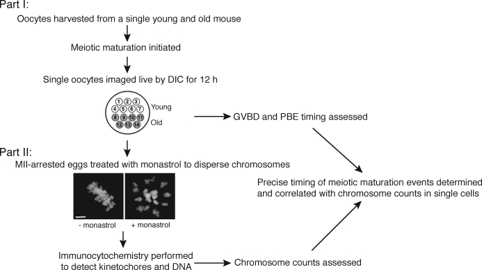
FIG. 1.
Experimental design for comparing meiotic progression and aneuploidy in individual young and old oocytes. Meiotic progression was monitored by time-lapse microscopy for 14 oocytes (seven young and seven old) in parallel (Part I). After live imaging, oocytes were cultured for 1 h in monastrol then fixed and processed for immunocytochemistry to label chromosomes and kinetochores (Part II). Chromosomes were counted to assess aneuploidy in each MII-arrested egg. The image shows that 1-h culture with monastrol effectively disperses the MII chromosomes, visualized by Sytox. Bar = 5 μm.

