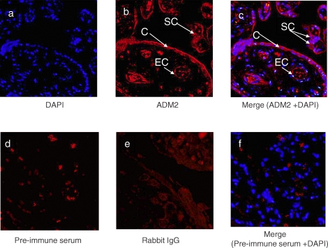
FIG. 2.
Single immunofluorescent staining for ADM2 in first-trimester villous trophoblastic tissue, followed by counterstaining with DAPI. a) Nuclei are stained blue when counterstained with DAPI. b) Red staining indicates ADM2 localization in cytotrophoblasts (C), syncytiotrophoblasts (SC), and endothelial cells (EC). c) Red staining indicates ADM2, and nuclei are stained blue when counterstained with DAPI in cytotrophoblasts, syncytiotrophoblasts, and endothelial cells. d) Negative control using preimmune serum. e) Negative control using rabbit IgG. f) Negative control staining using preimmune serum and counterstained with DAPI. Original magnification ×200.

